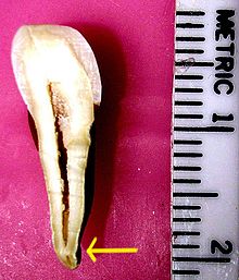
Apical foramen
The foramen apicale dentis (short form: foramen apicale , Latin : foramen 'opening', 'hole'; apicale 'towards the tip'; dentis 'of the tooth') is a term from the anatomy of the tooth . It describes the opening at the tip of the tooth through which the nerves , blood and lymph vessels enter the tooth and form the pulp . The rami alveolares (branching branches, arterioles and venules ) arise in the lower jaw from the inferior alveolar nerve or the inferior alveolar artery and the inferior alveolar vein , in the upper jaw from the superior alveolar nerve or the superior alveolar artery and superior alveolar vein . The entry of myelinated A-fibers , non- myelinated C-fibers and other nerves and lymph vessels occurs.
The anatomical root tip is referred to as the anatomical apex , the point on the tooth that is shown as the root tip in the X-ray image is referred to as the X-ray (radiological) apex . The distance from the foramen physiologicum to the foramen apicale is usually 0.5–1.0 mm, the distance from the foramen physiologicum to the radiological apex is 0.5–2.0 mm. The distances increase with age, as the formation of the apical root cement ( substantia ossea dentis ) increases. In many cases, the root canal gives off numerous accessory canals in the apical part, from which a so-called apical delta is created. At the apical constriction which begins periodontal ligament (ligament). The pulp tissue changes into a pulpo-periodontal mixed tissue .
In the event of tooth migration or tilting, the apical foramen itself can also "migrate" within a tooth, depending on how the function requires it. Dentin and cement are modified accordingly.
Therapeutic meaning
The apical foramen is of particular importance in the root canal treatment of pulpitic or necrotic teeth, because the root filling material should reach the apical foramen, but should not go beyond it. Ideally, root fillings should end at the apical constriction (foramen physiologicum) or just before it. In the case of teeth with a vital pulp, the healthy tissue beyond the constriction is prevented from being mechanically or chemically traumatized. In teeth with infected pulp, germs are prevented from spreading to the uninfected, periapical area (the area surrounding the tip of the root). If the constriction is expanded unnecessarily, there is also the risk of possible overfilling of the canal. The latter can lead to an undesirable tissue reaction (foreign body granuloma) and necessitate a root resection , with which the granuloma and the excess root filling material are surgically removed.
Individual evidence
- ↑ Michael Hülsmann: Endodontics . Georg Thieme Verlag, October 8, 2008, ISBN 978-3-13-156581-5 , p. 37.
- ↑ Elmar Hellwig, Joachim Klimek, Thomas Attin: Introduction to tooth preservation . Deutscher Ärzte-Verlag GmbH, 2013, ISBN 978-3-7691-3448-3 , p. 366.
- ^ Port, Euler, K. Greve: Textbook of dentistry . Springer-Verlag, March 8, 2013, ISBN 978-3-642-99128-8 , p. 99.


