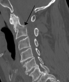
Head joint


As head joints are the joints between the base of the skull and the first cervical vertebra, the Atlas (atlanto-occipital joint) and the joints between the atlas and the second cervical vertebra, the axis designated (atlanto-axial joint). Together with the rest of the cervical spine, these joints cause the head to move in the three spatial planes : transversal (“turn”), coronal (“tilt”) and sagittal (“nod”). Colloquially, this upper area of the cervical spine is known as the "neck".
Upper head joint
The upper head joint or atlanto-occipital joint ( articulatio atlantooccipitalis ) lies between the two condyles of the occiput ( occiput ) and the fovea articularis cranialis of the atlas. It is an ellipsoidal joint that mainly enables extension and flexion, i.e. nodding movements (in English therefore also known as "Yes" joint , German "Yes" joint ). To a lesser extent, the head can also be tilted sideways.
The joint capsule is reinforced dorsally (towards the back) and ventrally (towards the abdomen) by membranes ( atlantooccipital membrane dorsalis and ventralis ). In the area of the dorsal membrane there is a larger hole between the two cervical vertebrae, which is only closed by this membrane. In this area it is possible with a cannula in the subarachnoid space or its extension ( cerebellomedullary cistern ) penetrate to a puncture of the cerebrospinal fluid (brain-spinal fluid, cerebrospinal fluid) to be performed. You can also use a sharp object to destroy the spinal cord there ("neck stitch"). In the spinal canal, the tectorial membrane runs over both head joints, and the atlantic cruciform ligament lies beneath it .
Lower head joint
The lower head joints or atlanto-axial joints ( articulatio atlantoaxialis ) are formed by the atlas and axis . There are the following joints:
- Articulatio atlantoaxialis mediana : The vertebral body of the axis is continued upwards ( cranially ) by a cone-shaped “tooth” ( dens axis ), which historically comes from the atlas. This tooth forms with its facies articularis anterior in the tooth pit of the atlas ( fovea dentis ) a so-called wheel or pivot joint ( articulatio trochoidea ). Furthermore, the dens axis articulates with its facies articularis posterior with the ligamentum transversum atlantis , which also secures it against backward movements. Interestingly, there are deposits of fiber cartilage cells on the surface of the ligament, which allow conclusions to be drawn about articulated contact with the dens axis. The ligament lies dorsal to the dens and is attached to the two lateral masses of the atlas.
- In the articulatio atlantoaxialis lateralis , the atlas and axis are connected via the lower and upper articular surfaces of the articular processes ( articular processes ).
These joint sections are enclosed by a common joint capsule and fixed by several additional straps . Around the dens of the axis predominantly rotational movements like the shake of the head (be "no joint" , "No" joint ) run. The pivot joint on the dens allows 20 ° –30 ° rotation to either side. About 70% of the head rotation occurs in this lower head joint, the rest in the rest of the cervical spine .
Interaction of the head joints
The head joints allow a very fine gradation of the movements of the head. By combining the nodding movements of the upper and the turning movements of the lower head joints, movements in all three spatial levels are possible.
Damage to the head joints
In the event of a fracture of the neck - a fracture of the tooth of the second cervical vertebra ( dens axis ) - or a tear in the ligaments of the dens axis , the elongated medulla ( medulla oblongata ) and the spinal cord can be severed or squeezed, which leads to the destruction of the respiratory and tract Circulatory center is coming. This results in immediate death, comparable to beheading . If an injured person without spontaneous breathing is suspected of having a fracture of the dens axis , the necessary intubation must be carried out with caution in the neutral position of the cervical spine in order to avoid possible or further damage to the extended marrow or spinal cord.

A missing or incomplete formation of the dens axis can be the cause of an atlanto-axial subluxation . This can trigger the same symptoms as a broken neck.
All alleged "instabilities" of the head joints that go hand in hand with no abnormalities in the ventral atlantodental joint are unproven claims that are of no relevance to conventional medicine and cannot cause any complaints.
The explanation for this is that the atlas is the 1st cervical vertebra, which is circular and rotates around the dens axis . This lies in the anterior section, which is why it articulates with the bony anterior portion of the atlas arch and forms the anterior atlantodental joint. The posterior articular surface of the dens axis articulates only with the cruciform ligamentum transversum atlantis , which is fixed to the right and left of the atlas, as well as up and down on the occiput and the 2nd cervical vertebrae ( fasciculus longitudinalis superior and inferior ). The dens axis is also suspended at its tip on the occiput and has two lateral wing-shaped ligaments ( lig. Alare ) that hold it in place. This ensures that the dens axis cannot press backwards on the spinal cord, except in the event of a break or when hanging on the gallows.
The ligaments cannot be seen or assessed on X-rays, so they are usually not mentioned in an X-ray report. There is no dorsal bony atlantodental joint, only a posterior articular surface of the dens axis, which is connected to the lig. transversum atlantis articulated. An X-ray and an MRI can be used to assess whether the position of the atlas and axis is correct and whether there are any malformations. This is the normal Dens ap image, and if the atlantodental joint is abnormal, an MRI of the cervical spine. If an inconspicuous atlantodental joint can be seen in these recordings, regardless of whether there is an asymmetry to the left or right, which is a common norm variant without medical relevance, there is no instability of the head joints.
literature
- J. Fanghänel, F. Pera, F. Anderhuber and a. (Ed.): Waldeyer Anatomie des Menschen . 17th completely revised edition. de Gruyter, Berlin / New York 2003, ISBN 3-11-016561-9 , chap. 8.2.4 Head joints , p. 640 ff .
Individual evidence
- ^ Definition of the neck in the dictionary ; Retrieved August 14, 2011
- ↑ a b distance, atlantodental. Retrieved June 29, 2019 .
- ↑ instability of the upper cervical spine. In: Spine Therapy Charité Berlin. Retrieved June 29, 2019 (German).
- ↑ Sample radiographs and associated diagnoses. University of Bern , accessed on June 29, 2019 .
- ↑ BNC / Professional Association of Resident Surgeons, Surgery, Proctology, Pediatric Surgery, Vascular Surgery, Hand Surgery. Retrieved June 29, 2019 .
- ^ Deutscher Ärzteverlag GmbH, editorial office of the Deutsches Ärzteblatt: The cervical spine and cervical market trauma: Neurological diagnosis and differential diagnosis. May 22, 1998. Retrieved June 29, 2019 .
- ↑ The cervical spine. Retrieved July 1, 2019 .