hoof
The hoof is a horn structure that encloses the tip of the toe in odd-toed ungulates . The term is commonly used for the toe end organ of horses , donkeys and zebras ; in the case of even-toed ungulates , one speaks of claws . Anatomically, the hoof corresponds to the human fingernail . The hoof is a modified outer skin in which the subcutaneous tissue is missing except in the area of the padding and the epidermis is severely cornified .
The horse's hoof is central to its health. He must bear the weight of the horse and cushion the impact of every step and jump in order to prevent permanent damage to the joints. Furthermore, it increases the blood flow to the end organ during exercise. The hoof essentially consists of the front, relatively rigid part, which ensures a firm footing, and the rear, more flexible part, which is primarily responsible for breaking the impact.
construction
skeleton
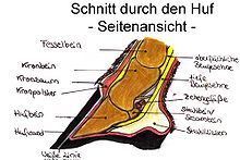
The coffin bone ( Os ungulare ), the navicular bone ( Os sesamoideum distale ) and the lower area of the coronary bone ( Os coronale ) belong to the skeleton of the toe end surrounded by the hoof .
Upholstery (subcutaneous tissue)
A subcutaneous tissue ( subcutis ) is not formed in most places or is identical to the periosteum . Only in a few places is it thickened to form cushions. They consist of soft fibrous connective tissue .
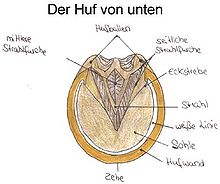
- The beam cushion or cushion lies between the two coffin cartilages with which it is connected by lateral adhesions. The frog fills the ball of the foot and, when the horse hits the ground, causes the cartilage to move upwards, thereby starting the hoof mechanism .
- The crown pad is located under the dermis in the area of the crown edge.
- The ball pad connects to the jet pad at the back and up and cushions the ball.
Dermis
The hoof dermis ensures, on the one hand, the connection to the coffin bone and thus the firm connection of the hoof capsule to the skeleton and, on the other hand, the growth of the hoof horn that forms the hoof capsule. Since the hoof horn has different functions in different parts of the hoof and therefore also has different structures, a distinction is made between the following six types of dermis.
Hemisphere, corium limbi
The hem dermis is the transition from the normal skin of the horse's leg to the coronet. It is only about five millimeters wide and the associated epidermis forms the relatively soft horn of the fringe band - this is the upper narrow edge of the hoof capsule - which is somewhat narrower than the adjacent hoof wall.
Kronlederhaut, Corium coronae
The corolla hide lies as a ring-shaped bead below the hem band and has villi a few millimeters long. The associated epidermis forms the horn of the hoof wall - this is the area of the hoof horn that surrounds the hoof on the side. Similar to the human fingernail, the hoof horn is continuously formed and pushed down to replace worn hoof horn. The growth rate is around eight to ten millimeters per month.
Wall dermis, Corium parietis
The dermis continues the coronet downwards and represents the largest part of the entire dermis in terms of surface area. It has an area of about one square meter per hoof and its leaflet structure ensures that the hoof wall is firmly held on the hoof. Like a Velcro fastener, it forms a movable connection that still guarantees an excellent hold.
Sole dermis , Corium soleare
The sole dermis in turn has short villi and covers the lower part of the coffin bone and ensures the growth of the hard part of the sole horn.
Radial dermis , Corium cunei
The frog dermis is responsible for the production of the elastic frog horn and also encloses the frog cushion.
Leather skin, corium tori
The leather skin forms the basis of the ball, the horn of which is soft and elastic.
Hoof capsule
The hoof capsule consists of the hoof wall , which laterally surrounds the hoof, the hoof sole, the hard part of the sole that closes it off from the ground and the hoof frog , the soft part of the sole. The bottom edge of the hoof wall, the so-called carrying edge , and the sole of the hoof are separated by the white line ( zona alba ), which also shows the farrier where to drive in nails without damaging the sensitive dermis. The upper edge of the hoof capsule is the coronet , which merges into the normal hairy skin via the narrow hem segment .
The hoof wall is divided into three areas from front to back. The front area is called the toe, the middle right and left is called the sidewall and the rear area is called the heel .
Hoof roll
As the navicular be navicular or its lower articulation surface, the deep flexor tendon and the navicular bursa ( navicular bursa ), respectively. Particularly when the hoof rolls, very heavy loads occur here. Diseases are common and occur almost exclusively on the forelimbs.
Hoof mechanism
The hoof mechanism is the elastic response of the hoof to stress. Contrary to the first impression one has of a horse's hoof, it is by no means rigid, but reacts flexibly to stress. When the hoof touches down, the front edge of the coronet is pulled back and down and the balls of the balls are pushed apart so that the frog also comes into contact with the ground and can thus pass on information about the soil condition to the nerve endings in the dermis. By contracting the balls of the feet when the pressure is relieved, an effect similar to that of a suction pump is triggered, which ensures good blood circulation in the hoof and, when the load is high, makes a decisive contribution to the overall blood circulation in the legs. That is why the four additional hearts of the horse are referred to when referring to the hooves.
Foal hoof
In order not to injure the mare's body with hard hooves, foals have a soft protective skin over the sole of the hoof, which only hardens in the first few days after birth and then expires over time. Since the hoof in the foal is also much narrower than it will be later in the adult horse, and the hoof does not grow in width, but can only grow downwards, you can always see edges on the hooves of young animals below which the hooves are narrower, as a growth spurt has started again and the coronet has expanded as a result.
Hoof care and protection
Before and after work, the horse 's hooves are scraped out during horse care . In order to avoid inflammation and pressure points, the hoof is regularly checked for foreign bodies, such as stones, and for its general condition. A healthy diet and a clean box are very important for the health of the hoof. Too much ammonia from urine in the litter, moisture and dirt often lead to thrush .
If the horse is used as a workhorse, this can lead to increased hoof abrasion, which can no longer be compensated for by natural regrowth. Then the hoof must be protected with a horseshoe , plastic shoe or hoof shoe . Nowadays, however, the typical leisure horse is usually so little stressed that artificial hoof protection is usually not necessary.
On a shod horse, the heels chafe on the iron due to the hoof mechanism and wear out, whereas the toe is fixed and does not wear out. This leads to a misalignment that must be corrected periodically. A shod horse therefore has to go to the farrier every 4 to 8 weeks. But even with an unshod horse, the hoof position must be checked periodically and corrected if necessary.


