Hydrops cochleae
| Classification according to ICD-10 | |
|---|---|
| H81.0 | Meniere's syndrome or dizziness |
| ICD-10 online (WHO version 2019) | |
Hydrops cochleae , endolymphatic hydrops or labyrinth hydrops describe a pathological increase ( hydrops ) of one of the fluids ( endolymph ) in the inner ear . It affects all chambers and channels filled with endolymph, both in the cochlea ( Latin : cochlea ) and in the organ of equilibrium, because both form a connected fluid system (Fig. 1). The chambers and channels that contain the endolymph are stretched in the event of a pathological increase in this fluid, which can lead to displacements and leaks of membranes and thus to hearing and balance disorders (dizziness).
For a long time, endolymphatic hydrops was only considered to be the direct cause ( pathomechanism ) of Meniere's disease . However, recent studies have shown that the hydrops cochleae can occur without the full picture of Menière's disease. It can therefore remain inconspicuous for a long time or permanently, or it can later develop into the trigger of Menière's disease. This knowledge is important for the timely diagnosis of Menière's disease in its early stage and the often possible preventive measures ( prophylaxis ) to avoid the full picture of the disease. The causes ( etiology ) of the endolymphatic hydrops - and thus also of Menière's disease - are still largely unclear.
history
Endolymphatic hydrops in human temporal bone preparations in Menière's disease was described for the first time in 1938 by Yamakawa in Japan and a few months later by Hallpike and Cairns in England. Both groups found a bulging of the Reissner membrane in the direction of the scala vestibuli and a concretion in the stria vascularis , in the aqueductus cochleae and in the internal auditory canal . Further confirmatory observations were made by Altmann and Fowler in 1943, by Lindsay in 1946, by Brunner in 1948, and by Paparella in 1984.
description
Hearing organ
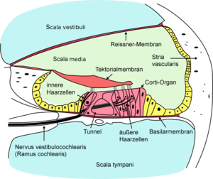
The cochlea of the inner ear has three passages (Fig. 2 & 3): atrial stairs ( scala vestibuli ), cochlea ( scala media or cochlear duct ) and tympanic stairs ( scala tympani ). The atrium staircase and the tympanic staircase are connected to one another via the helicotrema (Greek: snail's hole) at the "tip" of the cochlea, the snail thread ends at the tip of the snail. The cochlea is separated from the vestibule by the Reissner membrane and from the tympanic staircase by the basilar membrane . The organ of Corti is located in the cochlea , which converts sound waves into nerve impulses and thus the sensation of hearing. The three ducts are filled with two types of fluid of different composition, Scala vestibuli and Scala tympani with perilymph (blue in Fig. 3), the Scala media with endolymph (green in Fig. 3). The latter led to the name endolymphatic hydrops .
Under a cochlear hydrops ( hydrops = dropsy, abnormal fluid accumulation) is defined as an increase of the endolymph in the inner ear. With regard to the hearing organ (cochlea), this creates overpressure in the scala media . Its diameter increases, and the Reissner membrane is curved outwards in the direction of the vestibule (scala vestibuli). The immediate (proximate) causes are an overproduction of endolymph or insufficient drainage of the endolymph in the constant recycling process of this fluid. However, what leads to overproduction or congestion, i.e. the basic (ultimate) causes, is largely unclear.
It is assumed that too high a pressure on the Reissner membrane leads to temporary leaks or even ruptures of this membrane, with a subsequent local mixture of endolymph and perilymph. This can impair the chemical functionality of the fluids and the sensory cells affected by them, resulting in hearing impairments.
Balance organ
If this temporary, local mixture (poisoning) occurs in areas of the organ of equilibrium (Fig. 1, left half of the picture), this can lead to functional disorders of the sensory cells there and thus to attacks of dizziness, which are then referred to as Meniere's attacks .
root cause

The cause of endolymphatic hydrops is unclear. Explanatory attempts range from an excessive salt content in food intake, autoimmune reactions, allergic reactions to a viral disease. Since none of these possible causes could be sufficiently confirmed by clinical data as alone, it is assumed that in most cases a combination of several causes leads to an endolymphatic congestion. Since the micro-metabolism in the inner ear is highly complex and under the influence of the vegetative nervous system , stress and psychological stress are often mentioned as possible contributing or triggering factors. This coincides with the frequently observed personality image of the Menière patient, which was often characterized by a tendency towards ambition and perfectionism, i.e. the risk of putting oneself under excessive pressure.
Symptoms
The most common symptoms need not all appear. Often times, they come in groups such as dizziness without hearing loss and tinnitus, or hearing loss and tinnitus without dizziness.
- The patient can sometimes perceive the overpressure as a feeling that is similar to a dampening of the eardrum (e.g. from water in the ear canal ) or the middle ear (e.g. when the air pressure changes in an airplane). The English expression for this is " fullness of the ear ".
- Different types of hearing loss .
- Different types of sound sensation (tinnitus).
- Sometimes the disease also leads to a feeling of dizziness , which can occur in various forms and lengths of time.
In addition to other symptoms from the physical illness, psychological symptoms are often added after the diagnosis.
- Depression due to hearing loss and the resulting reduced quality of life.
- Fear of the disease getting worse and possible onset of Meniere's disease.
diagnosis
The following signs suggest the possibility of hydrops cochleae:
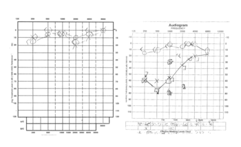
- Hearing test ( threshold audiogram ) shows hearing loss in the low and middle tone range, in contrast to the usual hearing loss due to noise damage, which mostly affects the high tone range.
- Hearing test ( discomfort threshold audiogram ) shows hypersensitivity ( intolerance ) to loud sound ( hyperacusis ).
- Information from the patient: If he describes a hearing disorder that mainly affects low frequencies, and "hearing through cotton wool" as well as low-frequency hum and pressure in the ear, hydrops cochleae must be considered alongside other causes such as sudden hearing loss or sound conduction disorder .
treatment
Betahistine
Endolymphatic hydrops is often treated with betahistine . However, as early as 2001, a systematic review (meta study) by the Cochrane Collaboration showed that there was insufficient data to be able to judge whether betahistine has any effect on Menière's disease at all.
In the case of hydrops, the situation is similar. After the expansion of the endolymph-filled vessels in the inner ear was quantitatively measurable in humans using magnetic resonance imaging (MRI) in recent years , a study with six patients showed an effect of betahistine in none of them and a case study of one patient over two years showed an end to it Spells of dizziness but worsening of hydrops and hearing in both ears.
Ventilation of the middle ear
The systems of pressure equalization in the inner ear are coupled in a highly complex way to the ongoing fluctuations in the static pressure in the middle ear and are stimulated by them. The details of the mechanisms have recently been largely clarified physiologically and anatomically .
The pressure regulation of the middle ear has a slow main component (gas exchange with middle ear tissue) and a fast secondary component (brief opening of the ear trumpet, the Eustachian tube ). The latter usually happens automatically as required, for example when chewing or yawning, but can also be brought about arbitrarily, for example when flying or diving.
In Menière patients, the pressure regulation of the middle ear is significantly worse than normal and there has been evidence since 1988 that additional ventilation of the middle ear can prevent attacks. In 1997 experimental evidence was obtained that additional ventilation of the middle ear actually counteracts the development of hydrops.
The method of choice for voluntary ventilation of the middle ear is the Valsalva method . It can be carried out almost anywhere spontaneously and without aids and is best combined with subsequent pressure equalization by yawning or chewing (slight cracking per ear). Valsalva, which should always be done carefully and with feeling, causes a brief increase in middle ear pressure, which then disappears again through the pressure equalization. This achieves an optimal effect on the aforementioned pressure regulation in the inner ear.
aftermath
Decreased hydrops can also cause permanent damage to the inner ear. A distinction must be made between objective and subjective problems.
Since the organ of Corti is stressed during the hydrops, tinnitus can remain even after the hydrops has receded. In addition, there may be an increased sensitivity to noise. Loud sound waves are perceived as annoying or almost painful. Hearing sensation disorders such as reverberation can also remain. There can also be a subjective feeling that the timbre of what is heard appears to be “remixed”. Some of these phenomena can be explained physically, such as permanent or temporary impairment of the organ of Corti , but also through psychological processes such as increased mental attention to the diseased ear.
All of these after-effects can decrease over time as the cochlea recovers and / or the brain adjusts to the new hearing sensation.
Differentiation from Menière's disease
Although Menière's disease is classified in the same way as endolymphatic hydrops in the classification according to ICD-10, it will probably have to be differentiated in the future. This results from the conclusions of the current research into the causes. More recent studies show that hydrops is probably not the result of a single cause and that fluctuations in the amount of endolymph can also occur in healthy people. The ear may react to all kinds of stress with endolymph congestion. The hearing organ, which evolved later in evolution, is probably more sensitive than the older equilibrium organ, which can better compensate for fluctuations in the endolymph. For Menière's disease, however, a specific cause may be assumed that leads to chronic or recurrent ( recurrent ) hydrops.
The most severe form of hydrops cochleae is a chronic hydrops or a recurrent hydrops including seizures with symptoms of Menière's triad. It takes an average of one year from the appearance of the first symptoms to the development of full Menière's disease. The practical experience of ear, nose and throat doctors and a study from Japan show that only about ten percent of diagnosed hydrops cochleae develop chronic Menière's disease. Approx. 70% of patients who do not develop Menière's disease develop normal hearing again. Only about 30% retain a fluctuating hearing ability, which could also be confirmed in follow-up examinations over ten years. From these statistical considerations, of course, it is not yet possible to conclusively draw conclusions about the patient's own clinical picture.
Individual evidence
- ↑ a b c d Olaf Michel: Menière's disease and related balance disorders . Georg Thieme Verlag, Stuttgart 1998, ISBN 3-13-104091-2 , p. 34 ff .
- ↑ a b c d e Helmut Schaaf (2007), Morbus Menière , Springer, Heidelberg, p. 58ff, ISBN 3-540-36960-0
- ^ Zenner HP, listening. Physiology, biochemistry, cell and neurobiology . Thieme, Stuttgart 1994, pp. 113-117.
- ↑ Health consultation ( Memento from October 25, 2007 in the Internet Archive )
- ^ CS Hallpike, JD Hood: Observations upon the neurological mechanism of the loudness recruitment phenomenon. In: Acta oto-laryngologica. Volume 50, 1959 Nov-Dec, ISSN 0001-6489 , pp. 472-486, PMID 14399131 .
- ↑ JD Hood, JP Poole: Tolerable limit of loudness: its clinical and physiological significance. In: The Journal of the Acoustical Society of America. Volume 40, Number 1, July 1966, ISSN 0001-4966 , pp. 47-53, PMID 5941765 .
- ^ MM Paparella, F. Mancini: Vestibular Meniere's disease. In: Otolaryngology - Head and Neck Surgery . Volume 93, Number 2, April 1985, ISSN 0194-5998 , pp. 148-151, PMID 3921902 .
- ↑ H. Levo, E. Kentala, J. Rasku, I. Pyykkö: Aural fullness in Menière's disease. In: Audiology & neuro-otology. Volume 19, number 6, 2014, ISSN 1421-9700 , pp. 395-399, doi : 10.1159 / 000363211 , PMID 25500936 .
- ↑ AL James, MJ Burton: Betahistine for Menière's disease or syndrome. In: The Cochrane database of systematic reviews. Number 1, 2001, ISSN 1469-493X , S. CD001873, doi : 10.1002 / 14651858.CD001873 , PMID 11279734 (Review).
- ↑ R. Gürkov, W. Flatz, D. Keeser, M. Strupp, B. Ertl-Wagner, E. Krause: Effect of standard-dose Betahistine on endolymphatic hydrops: an MRI study pilot. In: European archives of oto-rhino-laryngology: official journal of the European Federation of Oto-Rhino-Laryngological Societies (EUFOS): affiliated with the German Society for Oto-Rhino-Laryngology - Head and Neck Surgery. Volume 270, Number 4, March 2013, ISSN 1434-4726 , pp. 1231-1235, doi : 10.1007 / s00405-012-2087-3 , PMID 22760844 .
- ↑ C. Jerin, E. Krause, B. Ertl-Wagner, R. Gürkov: Longitudinal assessment of endolymphatic hydrops with contrast-enhanced magnetic resonance imaging of the labyrinth. In: Otology & neurotology: official publication of the American Otological Society, American Neurotology Society [and] European Academy of Otology and Neurotology. Volume 35, Number 5, June 2014, ISSN 1537-4505 , pp. 880-883, doi : 10.1097 / MAO.0000000000000393 , PMID 24770407 .
- ^ HP Wit, RA Feijen, FW Albers: Cochlear aqueduct flow resistance is not constant during evoked inner ear pressure change in the guinea pig. In: Hearing research. Volume 175, Number 1-2, January 2003, ISSN 0378-5955 , pp. 190-199, PMID 12527138 .
- ↑ R. Hofman, JM Segenhout, FW Albers, HP Wit: The relationship of the round window membrane to the cochlear aqueduct shown in three-dimensional imaging. In: Hearing research. Volume 209, Number 1-2, November 2005, ISSN 0378-5955 , pp. 19-23, doi : 10.1016 / j.heares.2005.06.004 , PMID 16039079 .
- ↑ M. Brattmo, B. Tideholm, B. Carlborg: Inadequate opening capacity of the eustachian tube in Meniere's disease. In: Acta oto-laryngologica. Volume 132, number 3, March 2012, ISSN 1651-2251 , pp. 255-260, doi : 10.3109 / 00016489.2011.637175 , PMID 22201512 .
- ^ P. Montandon, P. Guillemin, R. Häusler: Prevention of vertigo in Ménière's syndrome by means of transtympanic ventilation tubes. In: ORL; journal for oto-rhino-laryngology and its related specialties. Volume 50, Number 6, 1988, ISSN 0301-1569 , pp. 377-381, PMID 3231460 .
- ↑ RS Kimura, J. Hutta: Inhibition of experimentally induced endolymphatic hydrops by middle ear ventilation. In: European archives of oto-rhino-laryngology: official journal of the European Federation of Oto-Rhino-Laryngological Societies (EUFOS): affiliated with the German Society for Oto-Rhino-Laryngology - Head and Neck Surgery. Volume 254, Number 5, 1997, ISSN 0937-4477 , pp. 213-218, PMID 9195144 .


