Middle ear
The middle ear ( Latin : Auris media ) is a component of the human ear , but also of other terrestrial vertebrates .
It consists of a cavity that is on either side of the skull. This tympanic cavity ( Cavum tympani ) arises embryonically from the first pharynx pocket . It is filled with air and lined with a mucous membrane that is firmly attached to the periosteum . The middle ear is connected to the pharynx via the Eustachi tube ("ear trumpet") . It is used to equalize pressure with respect to the outside world.
Auditory ossicles

(2) hammer handle,
(3) stapes footplate with circle of the pilot laser,
(4) chorda tympani ,
(5) tendon of the stapes muscle (stapedius muscle)
In the middle ear are the tiny ossicles that connect the outer ear to the inner ear . The handle of the hammer ( malleus ) is fused with the eardrum ( membrana tympani ) . The hammer is articulated to the anvil ( incus connected), which in turn with the stirrup ( stapes is) in contact. The footplate of the stapes is movably fitted into the oval window ( fenestra ovalis or vestibularis ) of the inner ear ( auris interna ).
The ossicles are flexibly suspended by fine ligaments and muscles. They form a lever system that mechanically transfers the vibrations of the eardrum to the inner ear. Since the vibrations must be transferred from an air-filled space to a fluid-filled space (cochlea), the signal must be amplified. This signal amplification is achieved on the one hand by the leverage of the auditory ossicles, on the other hand by the fact that the eardrum has a larger area than the oval window. So there is an increase in sound pressure . This coupling is optimal in the range between 1 and 3 kHz, around 60% of the total sound power is transmitted from the eardrum to the inner ear. This mechanism is much less effective at lower frequencies, but also in the high frequency range.
Middle ear muscles
With the help of two small muscles, the properties of sound transmission can be changed: The tensor tympani muscle attaches to the hammer and tightens the eardrum. The stapedius muscle attaches to the stapes and tilts the stapes plate in the oval window. As a result, the coupling of the eardrum to the inner ear is worsened, the entire sound power is no longer transmitted to the inner ear, but part of it is reflected on the eardrum or diverted into the surrounding bones. In this way, the hearing can protect itself within certain limits from damage caused by excessive sound pressure .
Only the stapedius muscle is involved in the protective mechanism against high sound levels ; it contracts as a result of the stapedius reflex that is triggered by loud sound. The stapedius reflex starts at sound levels of 70 to 95 dB (stapedius reflex threshold) and is effective about 50 ms after the sound begins. The stapedius reflex acts on both ears, even if only one ear is exposed to a high sound level. By measuring the impedance of the external auditory canal , the use of the stapedius reflex can be observed and this can be used for diagnostic purposes.
phylogenesis

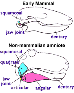
The temporomandibular joint (Latin: articulatio temporo-mandibularis ) is the flexible connection between the lower jaw and the rest of the skull . In vertebrates , more precisely from the jaws of the jaw (Gnathostomata), with the exception of mammals ( evolution of mammals ), the temporomandibular joint is formed by a connection between the articular bone and the quadratic bone ( primary jaw joint ).
In mammals ( therapsids , evolution of mammals ), these two bones have evolved into two of the three auditory ossicles and form the joint between the hammer and anvil. The temporomandibular joint in mammals is therefore a secondary temporomandibular joint and is formed by the articular bone of the lower jaw and the scaly part of the temporal bone and displaced into the cavum tympani .
Thus, the jawbones (articular, a small bone at the rear end of the lower jaw, and quadratum, a small bone at the rear end of the upper jaw) of the amniotes in mammals migrated into the middle ear. This enables better transmission of the acoustic signal. The earliest amniotes had a temporomandibular joint that was composed of the articular and the quadratum. All non-mammalian amniotes ( lizards , crocodiles , dinosaurs with their descendants, birds, and therapsids) are characterized by this jaw joint. However, mammals use a different temporomandibular joint system, which is composed only of the cover bones Dentale (the tooth-bearing lower jawbone) and Squamosum (small skull bone). In mammals, the quadratum and the articular became the anvil (incus) and the hammer (malleus) in the middle ear.
Diseases
An inflammation of the middle ear is called otitis media ( middle ear infection hereinafter).
See also
Web links
Individual evidence
- ↑ G. Djupesland: Electromyography of the tympanic muscles in man. In: Int. Audiol. 4, 33, 1965.
- ↑ K. Chiveralls, R. FitzSimons: Stapedial reflex action in normal subjects. In: Brit. J. Audiol. 7, 105, 1972.
- ↑ Pieter Nicolaas van Kampen: The tympanic region of the mammalian skull. W. Engelmann, Leipzig 1905.
- ↑ Johannes Müller, Linda A. Tsuji: Impedance-Matching Hearing in Paleozoic Reptiles: Evidence of Advanced Sensory Perception at an Early Stage of Amniote Evolution. at PLOS ONE
- ^ Mammalia: Overview - Palaeos
- ^ R. Cowen: History of Life . Blackwell Science, Oxford 2000, pp. 432 .
