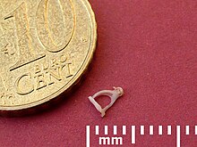
Auditory ossicles
The auditory bones ( Latin: ossicula auditūs , literally “bones of hearing”) are small bones in the middle ear of vertebrates with the exception of fish ( fish skull ), which transmit mechanical vibrations to the inner ear . The ossicles appear evolutionarily for the first time in amphibians in the form of a single ossicle , which is referred to as a column ( columella ) or stapes ( stapes ). In mammals, there are two additional ossicles, the hammer ( malleus ) and anvil ( incus ).
anatomy
Amphibians, reptiles and birds
In the bony fish (Osteichthyes) the two bones os quadratum and os articulare form the primary jaw joint. The hyomandibular bone, on the other hand, connects the quadratum bone with the bones of the skull. Also in the reptiles the os quadratum and os articulare form a primary temporomandibular joint, but here the os quadratum is directly connected to the roof of the skull. In amphibians, the hyomandibular bone became the columella and thus the ossicle. In contrast, in mammals, the lower jaw is directly connected to the temporal bone via a secondary jaw joint .
The stirrup ( stapes ), which in amphibians, reptiles and birds as little columns ( columella hereinafter) is an elongated bone rod and with its base ( base columellae ) in the oval window ( fenestra vestibuli ; also oval window, oval window ), a small opening between the middle and inner ear , anchored.
The opposite end of the columella has three cartilaginous extensions which Extracolumella ( Cartilago extracolumellaris ). In birds, amphibians and reptiles with an eardrum , the extracolumella is anchored to this. In animals with not hide or back educated middle ear cavity and lack the eardrum ( caecilians , salamanders , European spadefoot , toads , amphisbaenians and snakes ) is the Extracolumella (also plectrum called) attached to the skull. Here the auditory ossicle is not used to transmit sound waves, but rather ground vibrations that are transmitted via the skeleton to the oval window.
Mammals
The ossicles are the smallest bones in a mammal. The hammer weighs approx. 23 mg in humans, the anvil 27 mg and the stirrup approx. 2.5 mg. The ossicles are articulated to one another , fixed in the cavity of the middle ear by a ligament device and are covered in their entirety by the mucous membrane of the middle ear.
The hammer, the first bone in the chain of the ossicles, is fused with its spatula-shaped hammer handle ( Manubrium mallei ) with the eardrum ( Membrana tympani, Myrinx ). The end tendon of the tympanic membrane tensioner ( Musculus tensor tympani ) is attached to its muscular process ( processus muscularis ) . The head of the hammer ( caput mallei ) is connected to the body of the anvil ( corpus incudis ) in the hammer-anvil joint ( articulatio incudomallearis ).
On its long leg ( crus longum ), the anvil carries the lenticular process ( processus lenticularis ), which together with the stapes head ( caput stapedis ) forms the anvil-stapes joint ( articulatio incudostapedia ). The lens bone process is only connected to the anvil by a small bone bridge and connective tissue, so that it often breaks off during dissection and from the beginning of the 17th century, according to the first writer Franciscus Sylvius, was regarded as an independent ossicle (lens bone , os lenticulare , osselet de Sylvii ) . At the beginning of the 20th century, the lens bone was stripped of its status as an independent bone in human anatomy and zoology and this term was deleted from the human anatomical nomenclature . This term has been used in veterinary nomenclature to this day. An independent lens bone does not occur in any mammal.
The end tendon of the stapes muscle ( stapedius muscle ) attaches to the head of the stapes . The stapes plate ( base stapedis ) is anchored via the stapes ring ligament ( ligamentum annulare stapedis ) with a tape stick ( syndesmosis tympanostapedia ) in the oval window of the temporal bone pyramid .
histology
Histologically , the ossicles show significant differences to the other bones. They not only contain lamellar bones , but also woven bones , cartilage , calcified cartilage, so-called globuli ossei and interglobular spaces, as well as the so-called strand bone.
The strand bone is an embryonically formed bone substance, the collagen fibrils of which run in all levels of the room and intertwine to form strands similar to those of hair. The Globuli ossei are spherical collections of bone substance, on the edge of which cartilage remains persist. These cartilage remnants calcify and are called interglobular spaces .
Development history
The ear was originally not used to perceive sound, but as an organ of equilibrium. The columella is already at the beginning of the evolution of land vertebrates found on early amphibians. In sharks and bony fish, this bone is still part of the upper jaw suspension as a so-called hyomandibular . The homologue of the columella in mammals is the stapes, but only the part of the columella on the inner ear side, since the extra columella has receded in mammals. The other two bones were first identified as fossils in the mammalian species Hadrocodium wui from the Jurassic Age . In the other vertebrates they form the (primary) jaw joint as articular and quadratum , which in mammals is replaced by a secondary temporomandibular joint arising elsewhere during fetal development . Two of the exclusive features of mammals concern the auditory ossicles: only in representatives of this class are three auditory ossicles formed and only in these does the stapes have a stirrup-like shape.
The Reichert - Gaupp theory, which is currently still recognized by most scientists, was initially established on the basis of the positional relationships between the ossicles and the nerves of the middle ear. It assumes that the bones of the primary temporomandibular joint are included in the ossicular chain, which in turn arises from the cartilages of the first and second gill arches .
In the embryo , the hammer arises from the three bones articular , angular and gonial of the mandibular arch . The articular bone forms the main part of the hammer, the goniale its rostral process . The connection to the angular bone , which later develops into the tympanicum , loosens so that the ossicular chain can vibrate freely. In the human fetus, the hammer begins to ossify from a single ossification center in the 4th month , and in the 7th month the ossification is then almost completely completed.
According to the Reichert-Gaupp theory, the anvil also arises completely from the first gill arch ( mandibular arch , Meckel's cartilage ), although it is still not clear whether parts of the anvil also arise from the second gill arch ( hyoid arch ). The anvil is ossified in the same way as the hammer, the lenticular process in humans is not formed until the end of the 5th month.
According to the Reichert-Gaupp theory, the stirrup arises from the hyoid arch ( Reichert's cartilage ), although recent work suggests that its footplate arises from the ear capsule, i.e. it has two embryonic precursors. The shape of the stapes, unique in the animal kingdom, in mammals with two legs ( crura stapedis ) comes about because the stapes in the embryo develop around the later receding stapes artery ( arteria stapedia ). The stapes have two ossification centers, one in the middle of each leg. Ossification begins in the human fetus towards the end of the 4th month, around the end of the 8th month the stapes head and footplate are ossified. Thus, at the time of birth, the ossicles are fully grown and completely ossified.
The Reichert-Gaupp theory was repeatedly questioned. Otto currently suspects undetectable material shifts in the second gill arch, which should be the starting point for all three auditory ossicles, so excludes any involvement of the first gill arch. However, this hypothesis has so far been considered an unproven assumption.
function
In animals with an eardrum, the ossicles have the task of coupling the vibrations of the eardrum as optimally as possible to the inner ear and protecting the inner ear from excessive sound pressures.
An exception are the turtles , whose eardrum is too thick to swing. In animals without an eardrum, the columella only serves to transmit vibrations from the skeleton to the inner ear - these animals are largely deaf. The columella of reptiles mostly serves to transmit sound and is then a bone rod that is lightly built so as not to hinder this function. Some reptiles such as the marine reptiles (Ichthyosauria) and some Pelycosauriern (Pelycosauria) columella is however built much more massive and its function in the auditory apparatus unknown.
Coupling of the eardrum to the inner ear
In order to stimulate the fluid of the inner ear, known as perilymph , to vibrate, significantly higher pressures and significantly smaller deflections are required at the oval window than at the eardrum . Since air has a significantly lower acoustic impedance than a liquid, a direct coupling of the air oscillation would only give 2% of the sound power to the perilymph, the rest would be reflected .
The ossicles, together with the eardrum and the atrial membrane on the oval window, therefore serve as impedance converters . Low sound pressures and high deflections of the air in front of the eardrum are converted into high pressures and low deflections of the perilymph at the oval window ( Fenestra ovalis ) of the inner ear. The acoustic vibrations in the ear canal are converted into mechanical vibrations in the ossicles via the eardrum. The mechanical vibrations of the ossicles are converted into fluid vibrations of the perilymph by means of the basilar membrane .
The system is normally tuned in such a way that the mechanical impedance of the eardrum-ossicle-inner ear system corresponds almost exactly to the acoustic impedance of the auditory canal, so that a large part of the sound power is transferred to the ossicles. The same applies to the coupling of the auditory ossicles to the inner ear. Between the very low mechanical impedance on the eardrum and the very high mechanical impedance on the inner ear, the ossicles act as an "impedance transformer". For this purpose, the ossicles are designed as a lever system that converts low forces and high deflections (= low impedance) on the eardrum into high forces and low deflections (= high impedance) on the atrial membrane.
The adaptation of the eardrum vibrations to the properties of the inner ear is normally optimal. Almost all of the sound power that penetrates the ear canal is passed on to the inner ear. The force exerted from the eardrum to the oval window increases by about a factor of 90 and the pressure by about a factor of 22. That means: If the eardrum and the oval window were rigidly connected, the sound transmission would be almost 30 decibels worse, and quiet noises would not be more noticeable.
The area ratio between the eardrum and the atrial membrane in the oval window supports the impedance conversion. The relatively large area of the eardrum leads to relatively large forces on the ossicles. The relatively small area of the oval window converts the force of the ossicles into a relatively large mechanical pressure on the perilymph in the inner ear. The area ratio between the eardrum and atrial membrane in humans is approx. 64 mm 2 to 3.2 mm 2 , i.e. approx. 20: 1, but if only the effectively vibrating part of the eardrum is considered without the part restricted in movement by the wall of the ear canal, the ratio is approx. 14: 1, for domestic dogs the ratio is 27: 1. This is why dogs usually hear much better than humans.
Protective function
With the help of two small muscles, the degree of deflection of the ossicles can be changed.
The tensor tympani muscle attaches to the hammer and tightens the eardrum. It protects against excessive movements of the ossicles and eardrum, for example when sneezing.
The stapedius muscle attaches to the stapes and tilts the stapes plate in the oval window. This worsens the coupling between the eardrum and the inner ear. The deflections of the auditory ossicles decrease, the sound power is in part no longer passed on to the oval window but to the surrounding bones or reflected on the eardrum. As a result, the entire sound power no longer reaches the inner ear. If sound levels of more than 80 to 100 dB SPL occur, reflex muscle tension of the stapedius muscle ( stapedius reflex ) occurs. The stapedius reflex protects the sensitive hair cells of the inner ear from excessive sound pressures .
Since the stapedius reflex leads to a change in the impedance transformation and thus to changes in the impedance of the eardrum, it can be detected using acoustic impedance measurements in the ear canal.
Filter characteristics
Since the middle ear consists of both vibrating masses (auditory ossicles) and elasticities (stiffness of the eardrum, atrial membrane and the ossicular suspensions), the system acts as a mechanical filter. However, the system is coordinated in such a way that it does not impair hearing in the greater part of the listening area. Only at the edges of the hearing area do the filter characteristics of the middle ear have a more significant influence.
Diseases
The pathological hardening ( sclerosis ) of the membrane that anchors the stapes plate in the oval window is called otosclerosis . It leads to a slowly increasing hearing loss , as it severely impairs the transmission of vibrations from the ossicular chain to the inner ear. With the help of microsurgery , the almost immobile stapes are replaced by an artificial stapes (stapes prosthesis).
Web links
- cochlea.org: How do I hear? - The ear (website in four languages by the French Rémi Pujol about the ear, Engl./Franz./span./portug.)
Individual evidence
- ↑ Federative Committee on Anatomical Terminology (FCAT) (1998). Terminologia Anatomica . Stuttgart: Thieme.
- ↑ Graphic on the development of the temporomandibular joints and their derivatives ( Memento of the original from February 25, 2014 in the Internet Archive ) Info: The archive link was automatically inserted and not yet checked. Please check the original and archive link according to the instructions and then remove this notice.
- ↑ F.-V. Salomon: Textbook of Poultry Anatomy. Gustav Fischer, Jena 1993, ISBN 3-334-60403-9 .
- ↑ GC Kent: Comparative anatomy of vertebrates. 7th edition. Mosby St. Louis 1992, ISBN 0-8016-6237-0 .
- ↑ a b D. Drenckhahn: Hearing and balance system. In: A. Benninghoff, D. Drenckhahn (Ed.): Anatomie. Volume 2, 16th edition. Urban & Fischer, Munich 2004, ISBN 3-437-42350-9 .
- ↑ a b c U. Gille: Ohr, Auris. In: F.-V. Salomon, H. Geyer, U. Gille (ed.): Anatomy for veterinary medicine. Enke Verlag, Stuttgart 2004, ISBN 3-8304-1007-7 , pp. 612-621.
- ↑ J. Hyrtl: Comparative anatomical studies on the internal auditory organ of humans and mammals . Ehrlich, Prague 1845.
- ↑ F. Wustrow: About the arrangement and the course of the fibrils in the strand bones of the ossicles. In: Z. Laryngol. Rhinol. Otol. 35/1956, pp. 544-553. PMID 13393200 , ISSN 0044-3018 .
- ^ A b F. Wustrow: The bone formation in the ossicles. In: Z. Laryngol. Rhinol. Otol. 35/1956, pp. 487-498. PMID 13361363 , ISSN 0044-3018
- ↑ D. Starck: Comparative anatomy of the vertebrates . Springer-Verlag , Berlin / Heidelberg / New York 1978, ISBN 3-540-08889-X .
- ↑ G. Fleischer: Evolutionary principles of the mammalian middle ear . Habilitation thesis . University of Giessen , 1978.
- ↑ K. Hinrichsen: Humanembryology . Springer, Berlin 1993, ISBN 3-540-18983-1 .
- ^ A b R. O'Rahilly, F. Müller: Embryology and teratology of humans . Hans Huber Verlag, Bern 1999, ISBN 3-456-82821-7 .
- ^ A b J. R. Whyte, L. Gonzalez, A. Cisneros, C. Yus, A. Torres, R. Sarrat: Fetal development of the human tympanic ossicular chain articulations. In: Cells Tissues Organs . 171/2002, pp. 241-249. PMID 12169821 , ISSN 1422-6405
- ^ Y. Masuda, R. Saito, Y. Endo, Y. Kondo, Y. Ogura: Histological development of stapes footplate in human embryos. In: Acta Med Okayama. 32/1978, pp. 109-117. PMID 150197
- ↑ H. Otto: Two previously unknown displacement movements in the branchial region of the human seedling - shown in the ontogenesis of the outer and middle ear including the periauricular region as well as in the pathogenesis of their malformations (the error of the Reichert-Gaupp theory) . Dissertation. Humboldt University, Berlin 1981.
- ↑ H. Penzlin: Textbook of animal physiology. 6th edition. Spectrum Academic Publishing House, 2005, ISBN 3-8274-0666-8 .
- ↑ H.-P. Zenner: Listen. Physiology, biochemistry, cell and neurobiology. Georg Thieme Verlag, Stuttgart 1994.


