Fish skull

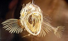
The skull of the fish is the oldest in the tribal history of the chordates (Chordata).
Components

The evolution of the fish skull can be traced back to three functionally different components - like the skull of the other, historically younger cranial or vertebrate animals.
Neurocranium
The neurocranium ("brain skull") protects (similar to the cephalopods ) the nerve center (brain) and, as far as possible, the large sense organs arranged here. As the lancet fish ( Branchiostoma ) shows, a skull is not necessary for a swimming or burrowing form without these sensory organs (the chorda dorsalis is sufficient to stiffen up to the tip of the body). As soon as the sensory organs ( nose , eyes , vestibular organ ) with the brain in between are present, however, the neurocranium becomes the axocranium , the fixed bow of an active swimming animal body. In fossil " bony fish " ( Palaeonisciformes , but also long before) the neurocranium consists of a single piece (albeit with cartilage indentations, probably against the risk of breakage), in recent bony fish it consists of 10 to 14 "cartilage or replacement bones" (because they are in embryonic cartilage skull caused by cartilage replacement); among the tetrapods , their number is slowly decreasing again.
Splanchnocranium
Since these " cranial animals " had gullet fissures from the beginning (primarily because of the consumption of food - see Tunicata ; the increasing size, however, soon brings with it the respiratory function: gill formation ), from a certain size skeletal supports are also necessary for these gill fissures : the splanchnocranium (from gr. σπλάγχνα pl "entrails"). This respiratory function is not necessarily tied to the front end ( cephalopods , crabs ), but proves to be advantageous "afterwards" - the Branchiocranium is (from the " Agnatha " [see Myxinoidea and Petromyzontida , in which it extends far behind the Neurocranium, while the dermatocranium is completely reduced] via the Chondrichthyes to the Osteichthyes - already with the Acanthodii ) tied more and more closely to the Axocranium (however, in the further development history towards air-breathing terrestrial vertebrates superfluous again and reduced to the hyoid bone and other "remnants"). The branchio- or splanchnocranium (the gill or pharyngeal arch with hyoid and jaw arch ) consists of about 33 named bones in bony fish, most of them in pairs and articulating to each other (in amphiarthroses ).
Dermatocranium

Later (?) Come - probably as a "reaction" to armored arthropods like the Ordovic Eurypterida ("sea scorpions") as enemies - on the front of the body (to protect soft tissues such as muscles, nerves, skin-sensory canals) (dozen) small bone plates of the skin also: the dermatocranium , which has very hard surface structures as a special feature, which could then differentiate themselves from the teeth in the area of the mouth opening . Cover bones also appear in the ectodermal cavity of the mouth and throat ( pharynx ), i. H. inside on the Branchiocranium, on (see also gill trap ). For decades there has been an argument as to whether the head lateral lines or the teeth represent the "origin" of the cover bones (both do not have to be). The cover bones and teeth also belong to the mesoderm - only the enamel is likely to be an ectodermal service. The “lime” skeleton in general can probably be “connected” to the sclerite plates (the stereoma ) of the echinodermata .
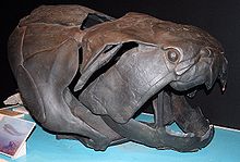
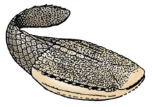
Today it is hardly possible to determine the sequence in which the three components appear, which, due to their weight, dictate different phylogenetic development tendencies. Even with the small Conodonta, only the teeth of the “skull” (?) Seem to have been preserved as hard parts of the living animal - everything else is sacrificed to the nectic way of life. In the thelodonti there is a thin skin ( scales ) armor. With the Ostracodermi u. a. however, the strong armor prevents life in the open water. The armored placodermi already had jaws, but no teeth of the appropriate size. They probably already had a swim bladder so that they could be the first great predator to roam through the open water despite heavy bone tissue ( Dunkleosteus was 8 m long). The “armor” ensures that the endocranium (inside - i.e. neuro- and splanchnocranium) can now remain gnarled (tender). Since the swim bladder also has its disadvantages, the " cartilaginous fish " did without it and part of the skeleton (bone tissue and dermatocranium) - but not the teeth.
Syncranium
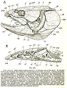
The recent (now living) bony fish have all three skull components (and at most secondary to little bones, no teeth and no swim bladder). In the sturgeon the skin bone armor is still quite independent, but in the higher forms (especially Teleostei ) there is a standardization (" syncranium "). The number of “bone individuals ” connected to one another by “sutures” ( sutures ; we know very little about the function of sutures) in synarthroses is also decreasing (through fusions or failures, as they say; for example, the dental is the Teleosteer a “fusion product” of Dentale, Mentomeckelianum, Spleniale, Surangulare (among others) at Amia and others, which however “failed” after being downsized in favor of the Dental, i.e. may have disappeared).
Fish skull bones
The head skeleton of a barbed fish , such as the perch ( Perca fluviatilis ), consists of almost 160 bones (for comparison: that of humans has 31). Most of them are paired (we only refer to the unpaired [u]). In the roof of the skull (i.e. in the dorsal view) we see the following cover bones (d, with sensory canal D) or cartilage bones (a) or mixed bones (a + d fused): Supraoccipitale (u, a, maybe also) d; D), supratemporal (D), d- parietal , d-epoticum, intercalare (d), frontal (D), nasal (D) and (mes-) ethmoideum (u, a, d).

The sides of the neurocranium make up the following ossifications: a-Exoccipitale, Opisthoticum (D), a-Prooticum, a-Sphenoticum (united with d-Postfrontale), a-Pteroticum (the three semicircular canals of the vestibular organ are located in the three otica) , Basisphenoid (u), Pterosphenoid, (Orbitosphenoid, u, only with thicker orbital septum), Ectetmoid (a with d-Praefrontale). The eye is protected by circumorbitalia (D; here 5 or 6 suborbitalia - the first is often called praeorbital or lacrimale). The gill cover (d) includes operculare, suboperculare and interoperculare. The ventral view shows the "base of the skull": Basioccipitale (u), Parasphenoid (u, d) and vomer (u, d).
The splanchnocranium consists of seven “visceral arches”, the traceability of which to “gill arches” is not guaranteed for all. Four real gill arches each consist (from top to bottom) of pharyngo-, epi-, cerato- and hypobranchial and one basibranchial each (u); the "5. Arch "consists only of the" Cerato element "(they say. Some elements remain cartilaginous throughout their life). These cartilage bones (a; s. Pharyngealia ) have tooth plates inside (d); a and d are grown together to very different degrees (the "5th Ceratobranchiale" is always mixed bone). The hyoid lies ventrally in front of the gill cage (the seven d-Branchiostegal radii are attached to it; see Branchiostegal apparatus ), consisting of epi-, cerato- and two hypohyal (n) and the basihyale (u, with d-glossohyale), dorsal the (a) Hyomandibulare, which is covered on the outside by the preoperculare (D). The hyoid is articulated to the quadratum by the rod-shaped inter- or stylohyal, the hyomandibular by the ("wedged") symplecticum . The onetime "arch" (now jockstrap called) consists of: Palatinum (at Percoidei only d) two (d) pterygoid (ectoderm and endoderm), Metapterygoid (a to d), Quadratum (a, d?) . The mostly wide cartilage zone of the jockstrap is functionally important. Three ( but ten for Amia calva !) Bones: Dentals (D, with a-part), articulars (a) and angulars, form the lower jaw , in which the elastic, cartilaginous mandible is always preserved. The " upper jaw " of the Teleostei, however, only consists of two cover bones (originally the nasal region): the premaxillary (in barbed fish only on the edge of the mouth, toothed, with (u) rostral cartilage) and maxillary (in more primitive fish still toothed). Finally, two tendon ossifications: the Urohyale (u, ventrally on the hyoid) and the Sesame Articular (attachment of the masseter tendon on the lower jaw).

In fish, the shoulder girdle can also functionally be counted as part of the skull (e.g. because it supports the gill cavity at the back), it consists of (d-) supracleithral, cleithral and the two a-bones coracoid and scapular ; it is guided by the post-temporal at the occiput (cf. the reverse semantics in Dunkleosteus [Fig.]).
Functional
(Here is only a very rough overview - the variety of the fish skull is vast; cf. Gregory 1933.) The skull is steered by the spine, with more primitive forms still by the chorda, which can extend into it up to the base phenoid. The articulation takes place on the basioccipital, often also on the exoccipitalia, and is usually not very mobile (bow effect!). Greater mobility is given z. B. by lifting the skull to snap (z. B. Zander ; Uranoscopus , excessive in some deep-sea fish such as Malacosteinae ); "Biting down" in Clariidae ; The salamander fish Lepidogalaxias has become famous for its lateral head movements (which is why it was even assumed that it can no longer move its eyes. Vertebrates have movable eyes - the cephalopods show that it can be done without). The purpose of elevating the skull is to align the direction of movement and the snap direction in free-swimming fish (because the lower jaw mainly opens downwards). Another possibility was to only lift the anterior skull so that the coordinating vestibular organ (in the "otoccipital block") could remain at rest (intracranial kinetics, only with sarcopterygii , recently Latimeria ; disadvantage: bow effect weakened!). There were also fish that could only lift the nasal capsule (recently with Lepidosiren and Cobitidae , but still unclear). The jockstrap and maxillary apparatus (see fish mouth ) of higher fish orders also help here.


Apart from the trunk muscles, about 60 muscles are attached to the skull (or parts of it) (most of them in pairs, of course); some of them divided into portions, e.g. B. the masticatory muscle in up to 12. The jockstrap enables the lateral expansion of the pharyngeal space (for breathing, sucking and swallowing); the hyomandibular on the pro, sphenoid and pteroticum and the palatineum on the mesoid and ectethmoid (but the ectopterygoid in carp and catfish). The movements of the jockstrap often take the lacrimal with them and thus serve to ventilate the olfactory organ. The lower jaw articulated on the quadratum (saddle joint! Diarthrosis ) has to take part in the transverse expansion in the symphysis and rotates lengthways with the teeth outwards. Long teeth can also be foldable (thanks to elastic ligaments they straighten up again after the prey passage; e.g. in Merlucciidae ). There are even fish with an intra- mandibular joint: to increase the bite force ( Scaridae ; "Streptognathy"). The movements of the hyoid and gill cover are equally important for breathing and food acquisition; z. B. Operkel-lifting is used in most fish, via the interoperculum, to open the mouth. The gill basket is "suspended" from the base of the skull (parasphenoid) by means of the pharyngobranchial I, in order to enable rapid looping movements of the dorsal pharyngealia (with gripping tooth plates) in the forms with retractor muscles . These pharyngealia are articulated pharyngo- and epibranchalia (II-IV), which interact with the mentioned "Ceratobranchialia V". For mollusc-eaters etc. Ä. ( Sparidae et al.) It even comes to the formation of a proper ball joint between the pharyngeal apparatus and the parasphenoid. In the Scaridae, the fused “Ceratobranchialia V” articulate inside the Cleithralia for increased stability.
Several functional achievements of the fish skull were passed on from the sarcopterygians to the tetrapods to the birds. It is only in the case of mammals that most things are no longer advantageous and are given up again (akinetic skull - similar to the Dipnoi ).
See also
literature
- William K. Gregory: Fish skulls. A study of the evolution of natural mechanisms. New York 1933, OCLC 251016995 . (1959 edition on archive.org)
- Wilhelm Harder : Head region and shoulder girdle of the fish. Swiss beard, Stuttgart / Berlin 1965.
- Wilhelm Marinelli , Anneliese Strenger: Comparative anatomy and morphology of vertebrates I-IV. Deuticke, Vienna 1953–1973.
- Wilfried Westheide , Reinhard Rieger (Hrsg.): Special zoology. Part 2: vertebrates. Gustav Fischer, Stuttgart / Jena / New York 2004.
Web links
- Compact Lexicon of Biology, Spectrum Academic Publishing House, Skull , Cranium


