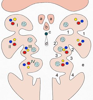
Gill arch
The gill arches , also called branchial arches ( Arcus branchiales ), throat arches , pharyngeal arches or visceral arches (southern German is usually used as plural 'arches'), are formations of the gill gut in vertebrates . The process of formation is known as branchiomerism . In the mammalian embryo , six such arcs are formed, with the fifth and sixth usually only being rudimentary .
Typical of the gill arches are their metameric structure, i.e. each gill arch has the same structure. Each branchial arch has a nucleus of mesoderm , from which a cartilage and muscle system later emerges, a branchial arch nerve from the neural crest and a branchial arch artery. The serial or metameric sequence of the branchial arch nerves has nothing in common with the segmental arrangement of the spinal nerves . The head and trunk regions are organized differently, in other words, the branching of the branchial arch and the metamerism of the abdominal wall are to be regarded as independent of one another.
In fish , the membranes between the furrow and pocket tear and the definitive gills appear . The existence of such gill arches and furrows in embryos of higher vertebrates was first described by Martin Rathke .
Gill arch
Each gill arch has a gill arch artery and - vein , a gill arch nerve and a muscle - and a cartilage model . In higher vertebrates, many organs develop from the gill arches , which are therefore referred to as branchiogenic organs .
First gill arch
From the first branchial arch ( mandibular arch large parts are formed of the face, such as) the upper jaw ( maxilla ), lower jaw ( mandible ) and the palate , as well as in mammals, the auditory ossicles hammer and anvil (but not the stirrup). In fish, amphibians, reptiles and birds, the bones of the primary temporomandibular joint develop instead of the ossicles: Os articulare and Os quadratum . The cartilage system is called Meckel's cartilage .
The masticatory muscles arise from the muscle application . The artery of the first branchial arch largely recedes, but is to a small extent involved in the formation of the external carotid and maxillary arteries . The first branchial arch nerve is part of the fifth cranial nerve , the trigeminal nerve .
Second gill arch
The second branchial arch is also known as the hyoid arch (or hyoid arch). The upper part of the hyoid bone (cornu minus; smaller part of the corpus), the processus styloidus of the temporal bone and the upper part of the cartilage also form the auditory ossicle of the stapes .
The mimic muscles , as well as the stapedius muscle , the stylohyoid muscle and the posterior part of the digastricus muscle, arise from the muscular system . The second branchial arch nerve is the seventh cranial nerve, the facial nerve . The associated artery initially develops into the arteria stapedia . This in turn recedes completely, so that only a vascular hole remains in the stapes.
Third gill arch
The third branchial arch cartilage develops to the lower part of the hyoid bone (cornu majus; larger part of the corpus), the muscular system to the musculus stylopharyngeus , which is accordingly innervated in adults by the third branchial arch nerve, the ninth cranial nerve glossopharyngeal nerve.
The artery of the third branchial arch develops together with the dorsal aorta to form the internal carotid artery .
Fourth gill arch
The fourth branchial arch and its cartilage transform into the upper part of the larynx . The external muscles of the larynx and a part of the throat muscles develop from the muscle system . The fourth branchial arch nerve is the superior laryngeal nerve , branch of the vagus nerve (tenth cranial nerve) that innervates the external muscles of the larynx (cricothyroid muscle).
The arteries of the fourth branchial arch develop differently on both sides: the aortic arch arises from the left artery, and the right subclavian artery.
Fifth gill arch
The fifth gill arch is only rudimentary or not at all. There are no definite structures.
Sixth gill arch
The sixth gill arch and its cartilage transforms into the lower part of the larynx. The muscle system develops accordingly to the internal larynx muscles . The 6th branchial arch nerve is the recurrent laryngeal nerve , a branch of the vagus nerve (tenth cranial nerve) that innervates the internal larynx muscles with the exception of the cricothyroid muscle.
The arteries of the sixth gill arch develop on both sides different: Links created the pulmonary trunk and the ductus arteriosus , right pulmonary artery .
Throat pockets
Between the six gill arches, five pharyngeal pouches or gill slits ( Sacci pharyngeales ) develop on the inside , from whose endoderm definitive structures also develop.
- From the first pharyngeal pouches caused recess tubotympanicus , from which the tympanic cavity and the Eustachian tube (eustachian tube) form. This also forms the endodermal part of the eardrum .
- The second pharynx forms the system for the palatine tonsil .
- The lower parathyroid glands ( glandula parathyroidea inferior ) and the thymus develop from the wall material of the third pharynx .
- The upper parathyroid glands ( glandula parathyroidea superior ) and - except in mammals - the ultimobranchial body form from the fourth pharynx .
- The C cells , which migrate into the thyroid gland in mammals, develop from the material of the fifth pharynx .

Gullet furrows
Externally, four pharyngeal or gill furrows ( sulci branchiales or ductus branchiales ) develop from the ectoderm . A definite structure emerges only from the first pharyngeal groove, namely the external auditory canal and the ectodermal part of the eardrum. The first pharyngeal furrow and the first pharynx grow towards each other until they are only separated by a fine skin, the eardrum .
Due to the strong growth of the second branchial arch, the second, third and fourth furrows are usually closed to form the cervical sinus . This obliterates. If this closure is incomplete, neck fistulas and cysts can form.
See also
literature
- Thomas W. Sadler: Medical Embryology. Normal human development and its malformations . 10th edition. Thieme, Stuttgart 2003, ISBN 3-13-446610-4 , p. 322-335 (English: Medical embryology . Translated by Ulrich Drews).
- Ulrike Bommas-Ebert, Philipp Teubner, Rainer Voß: Short textbook anatomy and embryology . 2nd Edition. Thieme, Stuttgart 2006, ISBN 3-13-135532-8 , p. 59 ff .
Web links
- Jubin Kashef, Almut Köhler, Doris Wedlich: The route planner of the neural crest cells. In: BIOspektrum March 2007, 13th year.
Individual evidence
- ↑ Jan Langmann: Medical Embryology. Normal human development and its malformations. Thieme, Stuttgart / New York 1980, ISBN 3-13-446606-6 , p. 265.
- ↑ Thomas W. Sadler: Pocket Textbook Embryology: The normal human development and its malformations . 12., revised. and exp. Ed. Thieme, Stuttgart 2014, ISBN 3-13-446612-0 .

