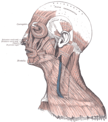
Facial muscles
The facial muscles ( Latin Musculi faciei , muscles of the face ) are a group of striated muscles that are responsible for facial expressions and thus for expressing emotions as well as individual facial expressions. They also have a protective function by closing head openings such as eyes and mouth. In addition, the facial muscles participate in language formation and food intake. In animals, they influence directional hearing and social behavior ("ear play") through the position of the auricles .
anatomy
The facial muscles form the superficial layer of the head muscles ( musculi capitis ). It arises from a uniform muscle plate ( panniculus carnosus ). This differentiates into a system of delicate muscles that either run circularly around or radially to the head openings (eye, ear, nose) and thus serve either to narrow or expand them. The ear muscles are used to move the auricle, but are not able to close the outer ear opening .
The characteristics of the facial muscles are:
- they advertise on the border with the epidermis
- they move the skin (not a joint, as in skeletal muscles ).
- Most of the facial muscles are not covered by a fascia (with the exception of the buccinator muscle )
- they are innervated by branches of the facial nerve (VII cranial nerve)
The paired facial muscles in humans include:
- The occipitofrontalis muscle (occipital-forehead muscle) moves the scalp . It can be subdivided into the venter frontalis (forehead-abdomen), also known as the eyebrow lifter ( musculus frontalis ), and the venter occipitalis (occiput-belly) or musculus occipitalis . The galea aponeurotica lies between the two .
- The temporoparietalis muscle (temple-vertex muscle) runs between the ear cartilage and the galea aponeurotica. With the occipitofrontalis muscle, it is combined to form the scalp muscles ( epicranium muscles ).
- The procerus muscle (slender muscle) pulls the meninges down between the eyebrows.
- The nasal muscle (nasal muscle ) can be divided into the nostril constrictor ( musculus compressor nasi , syn. Pars transversa ) and the nostril widener ( musculus dilatator naris , syn. Pars alaris ).
- The depressor septi nasi muscle (pulling down the nasal septum) pulls the nostrils down and narrows the nostril.
- The orbicularis oculi muscle (eye ring muscle) encloses the respective eye. It closes the eyelids and is responsible for the protective eyelid reflex and blinking.
- The corrugator supercilii muscle (eyebrow furrower) can pull the central side of the eyebrows down and thus causes the frown.
- The depressor supercilii muscle (pulling the eyebrow down) pulls the eyebrow down.
- The auricularis anterior muscle (anterior ear muscle) pulls the auricle slightly forward. In animals, a group of six anterior ear muscles is distinguished here.
- The superior auricular muscle (upper auricle) pulls the auricle upwards. In animal anatomy, a distinction is made between three upper auricular muscles, which are summarized as the auriculares dorsales muscles .
- The auricularis posterior muscle (rear ear muscle) pulls the auricle slightly backwards. In animal anatomy, a distinction is made between four rear ear muscles, which are summarized as the auriculares caudales muscles .
- The orbicularis oris muscle (oral ring muscle , also lip tendon muscle ) surrounds the mouth in a ring. It is not attached to any bone, but is held by other muscles. Therefore he is particularly agile.
- The depressor anguli oris muscle (corner puller) connects the lower lip at the corner of the mouth with the lower edge of the jaw.
- The transversus menti muscle (transverse chin muscle) runs between the two corner pullers on the chin.
- The musculus risorius (laughing muscle) extends from the corner of the mouth behind the jaw and pulls the corners of the mouth to the side.
- The zygomaticus major muscle (large zygomatic bone muscle), like the risorius muscle, belongs to the laughing muscles and connects the zygomatic arch with the corner of the mouth.
- The zygomaticus minor muscle (small zygomatic muscle) connects the zygomatic arch with the lower lip.
- The levator labii superioris muscle (upper lip lifter ) pulls from below the eye socket to the upper lip , where it connects with the oral ring muscle.
- The levator labii superioris alaeque nasi muscle (upper lip lift of the nostril ) extends from the upper jaw to the upper lip and the nostril.
- The depressor labii inferioris muscle (lower lip puller) can pull the lower lip straight down.
- The levator anguli oris muscle pulls from the canine fossa to the corner of the mouth and lifts it up.
- The buccinator muscle (cheek or cheek muscle) forms the muscular basis of the cheeks .
- The mentalis muscle (chin muscle) attaches to the chin. If the skin on the chin looks wrinkled, this muscle is active.
The platysma is a flat neck muscle , which starts from the corner of the mouth and widens strongly over the neck to the base of the chest. Some authors do not count the platysma as part of the facial muscles because it is no longer in the face. The upper eyelid lifter ( musculus levator palpebrae superioris ) participates in facial expressions, but is counted among the eye muscles . It is not innervated by the facial nerve, but by the oculomotor nerve.
Facial Action Coding System
Movements that are generated by the facial muscles can be described and coded for different animal species with the help of the Facial Action Coding System (FACS) or systems derived from it.
Individual evidence
- ↑ a b Walther Graumann: Compact Textbook Anatomy . tape 2 . Schattauer, Stuttgart 2004, ISBN 978-3-7945-2062-6 , pp. 429 .
- ↑ Horst Erich König, Hans-Georg Liebich: Anatomy of domestic mammals: textbook and color atlas for study and practice . Schattauer, Stuttgart 2012, ISBN 978-3-7945-2832-5 , pp. 111 .
- ^ Johannes W. Rohen, Elke Lütjen-Drecoll: Functional anatomy of humans: textbook of macroscopic anatomy according to functional aspects . Schattauer, Stuttgart 2006, ISBN 978-3-7945-2440-2 , p. 89-91 .
- ↑ Wolfgang Dauber: Feneis' picture lexicon of anatomy . Georg Thieme, Stuttgart 2005, ISBN 978-3-13-330109-1 , p. 94-95 .
- ↑ a b c Uwe Gille: Ear, Auris. In: Franz-Viktor Salomon et al. (Ed.): Anatomy for veterinary medicine . Enke, Stuttgart 2004, ISBN 3-8304-1007-7 , pp. 612-621.
- ↑ Wolfgang Dauber: Feneis' picture lexicon of anatomy . Georg Thieme, Stuttgart 2005, ISBN 978-3-13-330109-1 , p. 98 .
- ↑ Wolfgang Dauber: Feneis' picture lexicon of anatomy . Georg Thieme, Stuttgart 2005, ISBN 978-3-13-330109-1 , p. 444 .
