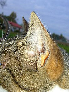
auricle
The auricle ( auricula auris ) is a part of the outer ear in mammals around the external auditory canal . It is shaped and stiff by a skin- covered structure of elastic cartilage ( Cartilago auriculae ). It is used to collect sound waves and localize a sound source . The auricles of most mammals are movable and can be changed in shape and position by the ear muscles , in humans only to a small extent.
The auricle is richly structured and shows protrusions and depressions with individually variable characteristics. The curved outer edge is formed by the ear strip ( helix ), curled up to varying degrees and sometimes with a pointed hump ( tuberculum auriculae ). The counter-ridge ( anthelix ) with two legs runs roughly parallel on the inside . It delimits the auricular cavity ( Cavum conchae ), which forms the access to the external auditory canal. The overlying hump ( tragus ) represents a movable ear cover in bats . In humans, the auricle has a cartilage-free appendage, the earlobe ( lobulus auriculae ).
Anatomy of the auricle of man
The auricles of humans are individually shaped. The shape of the auricle is inherited and can be used for proof of paternity. The mobility of the auricles is mediated by the ear muscles , but is of no functional significance in humans and at most allows for wobbling. The auricle consists of elastic cartilage, cartilage skin ( perichondrium ) and skin. The auricle is attached to the periosteum of the temporal bone and its mastoid process ( processus mastoideus ) by connective tissue . It is partially reinforced like a ribbon ( ligamentum auriculae anterius , posterius and superius ). The blood supply is provided by the arteriae auriculares anteriores and the branch auricularis of the arteria auricularis posterior . The blood outflow is realized through the veins of the same name and the superficial temporal veins . The lymph drainage takes place via the Lnn. retroauriculares and the Lnn. parotid .
The auricle has several elevations and depressions on the front and back. An elevation on one side corresponds to a corresponding depression on the other side. The outer edge is called the ear strip ( helix ). It sometimes shows a thickening, the tuberculum auriculae , also known as Darwin's ear cusp . Opposite it is the bulging counter-ridge ( anthelix ), which branches in the upper third of the ear into an upper and a lower leg ( crus superius anthelicis and crus inferius anthelicis ). The two legs border a triangular depression ( Fossa triangularis ). Between the helix and the anthelix there is a groove called a scapha . The ear cavity ( concha auriculae ) on the front side leads via the auditory canal entrance funnel ( cavum conchae ) to the external auditory canal . The hump that overlaps the entrance to the ear canal is known as the “billy goat” ( tragus ). The ear lobe (Latin lobulus auriculae ) is the soft part of the lower auricle that is not supported by the ear cartilage.
The most important depression on the back of the auricle is the retroauricular sulcus ( fossa anthelicis ) that runs from top to bottom and over almost the entire length of the ear . This depression corresponds to the back of the anhelix and can sometimes be quite pronounced.
The sound is refracted at the relief edges of the auricle and thus - depending on its frequency components - attenuated differently. The auricle ensures that the sound coming from behind is somewhat attenuated. This enables the brain to obtain information about the spatial origin of a sound source, in particular whether a sound comes from the front or the rear. The shape, size and position of the auricles are also important for the overall visual impression of the face.
Comparative anatomy

The auricle is diverse in mammals. Often it is pointed and is therefore also called pinna with the Latin word for feather, wing or fin . It is movable by various outer ear muscles , so that it can be used for sound location without moving the head. In addition, movements of the auricle also play a role in social communication ("ear play"). The skin is well supplied with blood and also contributes to heat dissipation in animals in warm climates. Therefore, the auricle is often relatively large in these animals, while it is very small in animals in colder climates. The elastic ear cartilage ( cartilago auriculae ) determines the shape and stiffness of the auricle.
The concave side of the auricle is referred to as the cone cavity ( scapha ), the outside as the dorsum of the ear ( dorsum auriculae ). The free edge is called the helix . The cone cavity leads into the auricular cone ( Cavum conchae ) and this in turn leads into the vertical part of the external auditory canal . Transferring the terms from the anatomy of the human ear, the front and inward edge of the entrance of the cavum conchae is referred to as the tragus , the opposite bulges anteriorly and laterally as the antitragus .
Development history
The auricle develops from the tissue around the first gill groove . The first three mesenchymal cusps of the first gill arch and the fourth to sixth mesenchymal cusps of the second gill arch form the actual auricle, while the concha and external auditory canal arise from the first gill groove. Around the seventh week of pregnancy, a large part of the mesenchyme of the first gill arch regresses, so that about 85% of the final auricle comes from the second gill arch.
Diseases
There are numerous malformations of the auricle that go as far as the complete absence of the same ( anotia ). In the case of malformations of grade I , all basic structures are in place. If the antelix is too weak or not developed at all, this causes the auricle to protrude ( apostasis otum ). Ear enlargements ( macrotia ) and cup deformities also belong to this group. Grade II malformations include cup ears and mini ears . Here the surgical effort for correction is already higher because additional cartilage and skin tissue is required for correction. In grade III malformations , the structures of the normal auricle are completely absent. They are often associated with malformations of the external auditory canal or the middle ear .
In an othematoma , there is bleeding between the ear cartilage and the outer skin.
Auricles and dummy head stereophony
In artificial head recording technology (binaural sound recording), microphones are built into a simulated head with auricles in the place of the ear canal (not the eardrum ) in order to give the listener a sound experience via headphones that is as true to the original as possible. If there are major differences between the auricles of the artificial head and one's own auricles, there are problems with the directional localization of the artificial head presentation. The front directions in particular appear inclined upwards by up to 30 ° (elevation) or can only be located at the rear. When listening with headphones , the ear cups are "switched off".
Web links
Individual evidence
- ^ A b Herbert Lippert: Anatomy on the living . Springer, Berlin 2013, ISBN 978-3-662-00661-0 , pp. 230 .
- ↑ Walther Graumann, Dieter Sasse: Compact textbook anatomy . tape 4 . Schattauer, 2005, ISBN 978-3-7945-2064-0 , pp. 98 .
- ↑ a b Uwe Gille: Ear, Auris . In: Franz-Viktor Salomon (Ed.): Anatomy for veterinary medicine . 2nd Edition. Enke, Stuttgart 2008, ISBN 978-3-8304-1075-1 , pp. 612-621 .
- ↑ Hilko Weerda: Surgery of the auricle: injuries, defects, anomalies . Thieme, Stuttgart 2004, ISBN 978-3-13-130181-9 , p. 105 .
- ↑ Hans-Peter Zenner: Practical therapy of ENT diseases: operating principles, conservative therapy, chemo- and radiochemotherapy, drug therapy, physical therapy, rehabilitation, psychosocial aftercare . Schattauer, 2008, ISBN 978-3-7945-2264-4 , pp. 83-84 .
- ↑ H. Weerda: Oto-Rhino-Laryngology in clinics and practices. Volume 1, Thieme 1994, pp. 511-512.

