Occiput

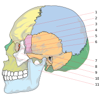
1. frontal bone (frontal) (yellow)
2. parietal (parietal) (light blue)
3. nose (nasal bone) (green)
4. ethmoid (ethmoid) (red)
5. lacrimal (lacrimal ) (light purple)
6. Sphenoid bone ( sphenoid bone ) (purple)
7. Occipital bone (occipital bone) (green)
8. Temporal bone (orange)
9. Zygomatic bone (white)
10. Upper jaw (maxilla) (yellow)
11. Lower jaw ( mandible ) (light blue)
The occiput (also occiput ; Latin os occipitale or occiput for short ) is the part of the brain skull located at the junction of the neck . It forms the posterior end of the cranial cavity and, with the atlas, the first head joint .
The occiput can be divided into three parts:
- Pars basilaris : floor part, part of the posterior base of the skull
- Pars lateralis : lateral part
-
Squama occipitalis : occipital scale (back), in which a distinction is made in the development phase between:
- Lower scale (caused by chondral ossification )
- Upper scale (caused by desmal ossification)
The sutura mendosa runs between the upper and lower scales . It ossifies in the 3rd month of life and is then visible as the Linea nuchae superior (upper neck line) on the bone.
The occiput is formed by the fusion of four bones, namely the basal, the two lateral and the upper occiput. The horizontal part is pierced by a hole as thick as a thumb (occipital hole or foramen magnum ), through which the spinal cord enters the vertebral canal from the cranial cavity , while the vertebral arteries enter the cranial cavity from outside. On either side of this hole are the two convex articular processes, by means of which the whole head can move, bend and stretch forward and backward on the first cervical vertebra.

On the underside, the occiput has an opening for the 12th cranial nerve ( nervus hypoglossus ), which is called the canalis nervi hypoglossi . In front of it rises an appendage, the processus jugularis , which in domestic animals carries the processus paracondylaris (origin of the musculus digastricus ).
The protuberantia occipitalis externa , to which muscles attach, and in horses and cattle also the neck band, rises on the neck surface .
literature
- Franz-Viktor Salomon: Bony skeleton . In: Franz-Viktor Salomon et al. (Hrsg.): Anatomie für die Tiermedizin . 3. Edition. Enke, Stuttgart 2015, ISBN 978-3-8304-1288-5 , pp. 92 .
- Meyers Konversations-Lexikon, 1888; Author collective, Verlag des Bibliographisches Institut, Leipzig and Vienna, fourth edition, 1885–1892; Volume 14: Rüböl - Sodawasser, page 372
Web links
Individual evidence
- ↑ Federative Committee on Anatomical Terminology (FCAT) (1998). Terminologia Anatomica . Stuttgart: Thieme


