Isocortex and Allocortex
Isocortex and allocortex ( Greek ίσος isos , German 'equal' άλλος allos , German 'different' and Latin cortex 'cortex' ) are the areas of the cerebral cortex that are defined and distinguished from one another according to histological criteria . The classification of the isocortex corresponds to the research results of Korbinian Brodmann , the couple Oskar Vogt and Cécile Vogt and the work of Constantin von Economo .
history
Karl Kleist (1879–1960), a student of Carl Wernicke (1848–1905), designed the first, much discussed map of the cerebral cortex .
Conceptual and methodological delimitations

From the histological differentiation into isocortex and allocortex, the developmental division of the cortex into neocortex , archicortex and palaeocortex is in principle to be distinguished from a methodological point of view. The answer to the question of whether there are any relationships between histological and developmental criteria is reserved for further research.
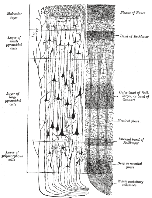
In humans, the majority (90%) of the cerebral cortex is made up of six well-defined layers, the so-called laminae . These differ in their cellular composition and in the course of the dominant pathways. The six-layer cortex is called the isocortex or homogenetic (identically structured) cortex. Some cerebral sections of the cerebrum, especially the olfactory brain and the hippocampus , are historically old and are characterized by a smaller number of layers, usually two or three laminae . These areas are called the allocortex, or heterogeneous (that is, "differently structured") cortex.
Layers of the isocortex
According to Ariëns Kappers , the six-layer type of bark fields shown below is said to have developed phylogenetically from a primitive three-layer structure. Such a three-layer structure is typical for the allocortex. However, objections have been raised to this promising and obvious theory.
Lamina I
-
Lamina molecularis (molecular layer):
- receives input from feedback neurons
- has no pyramidal cells
Lamina II
-
Lamina granularis externa (outer granular layer):
- receives input from feedback neurons
- is made up of small pyramidal cells, which give it a characteristic, granular appearance
Lamina III
-
Lamina pyramidalis externa (outer pyramidal layer ):
- is made up of pyramidal cells, which form cortico-cortical fiber connections with their axons
Lamina IV
-
Lamina granularis interna (inner granular layer):
- receives afferents from sensory neurons and is therefore very thick in the primary visual cortex or in the primary auditory cortex , but hardly pronounced in the motor cortex
- the inner granular layer is pervaded by strong bundles of horizontally running fibers, which in their entirety are referred to as the outer Baillarger stripe and come primarily from cortical afferents of numerous specific thalamic nuclei
- the inner granular layer is made up of partially heavily modified pyramidal cells with a star-shaped appearance and also of non-pyramidal cells and gets its name from those located here Granule cells
Lamina V
-
Lamina pyramidalis interna (inner pyramidal
layer ): - in the inner pyramidal layer there are the largest pyramidal cells, which with their axons form the main part of the cortex efferents to the deeper centers of the brain, for example to the basal ganglia, but not to the thalamus
- in the gyrus precentralis There are also some particularly large pyramidal cells, which are referred to there as Betz giant cells ; with their strong myelin sheaths, they form an essential part of the corticospinal tract (pyramidal tract)
- the cells of the inner pyramidal layer with their axons form the main exit system from the cortex
- including the inner pyramidal layer is traversed by a horizontally running strip of strongly myelinated fibers, which is referred to as the inner Baillarger strip , the inner Baillarger strip houses axon collaterals of neurons of the laminae II., III. and IV.
Lamina VI
-
Lamina multiformis (multiforme layer):
- the multiforme layer runs out into the underlying pith without sharp borders
- it contains, as the name suggests, in addition to smaller, morphologically different pyramidal
cells , also numerous non-pyramidal cells - the cells of the sixth layer of the Isocortex have afferents and efferents in other cortical layers or outside the cortex (extracortical), hardly any synapses are formed in lamina VI itself
- the pyramidal cells of the innermost layer of the isocortex direct their efferents primarily to the (specific) thalamic nuclei, the corticothalamic projections thus start from lamina VI, but the thalamocortical tracts end in layer IV
Physiology of the cerebral cortex
The structures listed above in no way allow an understandable insight into the physiology of the neo- and allocortex in terms of functional anatomy . As is well known, this only arises from a combination of structural, systematic and topological aspects.
Cell types
When shown with Golgi staining, the cerebral cortex generally contains 5 different types of neurons which, apart from their distribution in the various layers, suggest different functions.
Layers of the Allocortex
The Allocortex includes the formations of the Palaeopallium and the Archipallium such. B. the dentate gyrus with the exception of the cingulate gyrus , which belongs to the isocortex - cf. also the above-mentioned formations of the olfactory brain and the hippocampus. The names archicortex and palaeocortex have become established as the phylogenetically old structures belonging to the allocortex . The allocortex has the following layers:
- Lamina molecularis (Stratum molecularis): apical dendrites of the pyramidal cells
- Lamina pyramidalis (Stratum pyramidale): Cell bodies of the pyramidal cells
- Lamina multiformis (stratum oriens): basal dendrites of the pyramidal cells
See also
Individual evidence
- ↑ a b Alfred Benninghoff u. a .: Textbook of human anatomy. Shown with preference given to functional relationships. 3rd volume nervous system, skin and sensory organs. Urban and Schwarzenberg, Munich 1964, p. 231.
- ↑ Norbert Boss (Ed.): Roche Lexicon Medicine. 2nd Edition. Hoffmann-La Roche AG and Urban & Schwarzenberg, Munich 1987, ISBN 3-541-13191-8 , Stw.Allocortex, p. 48.
