Motor cortex
The Moto (r) cortex (from the Latin motor “mover”; from the Latin cortex “cortex”), also motor or somatomotor cortex, is a histologically delimited area of the cerebral cortex ( neocortex ) and the functional system from which voluntary movements are controlled and complex sequences can be put together from simple movement patterns. It forms the superordinate control unit of the pyramidal system and is located in the rear ( posterior ) zones of the frontal lobe .
Reflex movements ( internal muscle reflexes and external reflexes ), on the other hand, arise at a lower level (in the spinal cord or in the brain stem ) and are therefore not deliberately influenced. Other functional systems are involved in motor performance: For the control of muscle tone are basal ganglia important. The cooperation of the cerebellum is necessary for the spatial dimensioning, estimation of the necessary strength and speed and smoothing of the movements . Like the olive and the ruber nucleus, these are elements of the extrapyramidal motor system . This categorical classification (pyramidal-extrapyramidal) has since been abandoned because the cortical and subcortical systems largely overlap, and also non-motor brain areas, such as the rear (posterior) parietal cortex, play a crucial role in planning and performing easier and complex motor actions (e.g. purposeful gripping of objects).
Definition and anatomical containment
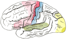
In the past, the entirety of the excitable cortex was viewed as the motor cortex. This was understood to be the sum of the cerebral areas, the external electrical stimulation of which can cause visible movements. Since this is also possible in practically all associative and some sensitive areas with sufficiently high stimulus voltages, there is a tendency today to include under the motor cortex only the areas in front of the central sulcus ( sulcus centralis ) that have a typical cytoarchitectonic structure.
These include the precentral gyrus and the posterior (posterior) parts of the superior , frontal medius and inferior gyri , as well as the anterior (anterior) section of the paracentral lobule .
histology
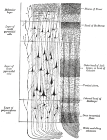
Histologically, the motor cortex belongs to the isocortex . This means that it has a defined six-layer basic structure that it shares with all phylogenetically young areas of the cerebral cortex. The inner granule cell layer, which is very pronounced in sensory areas and also occurs in prefrontal regions , is missing here or cannot be separated from the outer pyramidal cell layer . This is why one speaks of the agranular cortex . Due to the giant neurons in the 5th layer ( Betz giant cells ) that only occur here , the primary motor cortex is also referred to as the gigantocellular area. The frontally adjacent fields have a similar structure, but have no giant neurons, which is why they are also grouped together as the area paragigantocellularis . In some areas ( lobulus paracentralis ) the primary motor cortex reaches the greatest width of the cortex with 3 to 5 mm.
The individual layers are called:
- I. Lamina molecularis (molecular layer)
- II. Lamina granularis externa (outer granular layer)
- III. Pyramidalis lamina externa (external pyramidal cell layer)
- (IV. Lamina granularis interna ; inner granular cell layer)
- This layer is not histologically visible in the motor cortex. Basically, it is a pure question of definition whether one rejects their existence there or whether they are classified as III. Layer looks fused.
- V. lamina pyramidalis interna or lamina ganglionaris (inner pyramidal cell layer )
- This is where the pyramidal cells are located , which finally pass on the movement instructions through their cell processes ( axons ) to peripheral motor neurons. These include the so-called Betz giant cells in the primary motor cortex , which are among the largest cells in the human organism. However, they are clearly in the minority.
- VI. Lamina multiformis (multiform layer)
Functional and histological classification
Functionally, which is primarily-motor cortex (in Anglo-Saxon literature: M1 ) from the motor supplementary cortex ( supplementary motor area , SMA ) and premotor cortex ( premotor area , PMA or even PM ) distinguished. According to today's understanding, the latter serve to create certain sequences of movements from a learned fund of movements and to prepare voluntary (both conscious and unconscious) movements. The lying in the primary motor cortex motor neurons are the major common output of the motor cortex, as especially their axons the spinal cord and the motor cranial nerve nuclei reach. After switching to the peripheral motor neuron (anterior horn cell), the commands finally reach the voluntary muscles .
According to Korbinian Brodmann's histological brain atlas (see Brodmann area ), area 4 corresponds to the primary motor cortex; the supplementary motor cortex and the premotor cortex are formed by area 6 . Modern subdivisions, which are more function-oriented, differentiate a total of seven to nine fields, whereby these subdivisions are only based on studies on non-human primates, e.g. B. Rhesus monkeys have taken place (e.g. according to G. Rizzolatti F1 [= M1] to F7).
Primary motor cortex (M1)
Most of the primary motor cortex lies on the cortex in front of the central furrow ( gyrus precentralis ). What is remarkable is the so-called somatotopia , which means that neighboring regions of the body are also side by side in their representations on the primary motor cortex. The body is thus reduced in size and depicted upside down as a " homunculus " on the cerebral cortex. However, the proportions of the homunculus are distorted, as certain areas of the body have very finely tuned motor skills ; in humans this applies above all to the hand and the speech muscles . Other regions, on the other hand, can only be moved relatively roughly ( back ) or have a higher proportion of automatic regulation (holding and support muscles). The respective bark areas are correspondingly larger or smaller. However, the somatotopia in M1 is still much more pronounced than that of the primary sensory cortex (S1, area 3b), which has a precise representation of the body surface.
The descending ( efferent ) pathways that leave the cerebral cortex together form the corticonuclear tract , which supplies the motor cranial nerve nuclei, and the corticospinal tract, i.e. the pyramidal tract .
Premotor Bark (PMA / PM / PMC)
Quite large beef area ( Brodmann areae 6 and 8, = areae extrapyramidal) is located in front of the primary motor cortex and is located slightly lateral ( laterally ) on the convexity of the brain surface, while the supplementary-motor cortex to the center of the head to ( medial ) and predominantly on the other side of the mantle edge , so to speak on the opposite surfaces of the cerebral hemispheres. The task of this field is to create movement drafts and to coordinate them with the cerebellum and the basal ganglia . Sensory information is also included, which defines the necessary extent of movement, for example. An important subdivision of the premotor cortex is the dorsal premotor cortex (dPMC or PMd) and the ventral premotor cortex (vPMC or PMv), which is located somewhat deeper towards the Sylvian fissure. Both cortices are particularly involved in the transformation of visual information (e.g. position and shape of an object) into motor programs. Neurons of the dPMC code for the position of an object in space, whereas the vPMC controls the opening of the hand and finger for gripping an object. Both areas have intensive connections to the parietal cortex, with dPMC receiving projections from the superior parietal lobule (SPL) and vPMC from the inferior parietal lobule (IPL) and in particular the intraparietal sulcus (IPS). The parietal areas, on the other hand, are strongly connected to the visual cortical areas. Based on these anatomical facts, Rizzolatti developed a theory for cortical control of grasping movements, namely that visual information from the primary visual cortex (V1) reaches the parietal cortex via higher visual areas (V2-V8), in which the information is analyzed for location and shape which is passed on to the corresponding premotor areas that ultimately control the primary motor cortex. The examination of this concept is the subject of current brain research.
Some nerve cells in the premotor area are active during the planning and execution as well as the passive observation of the same movement in another individual (so-called mirror neurons ). One suspects their importance in imitative learning processes ( see also: learning on the model ). However, more recent hypotheses tend to assume that the mirror neurons (which can also be found in other brain areas such as the inferior parietal lobule) rather encode the “understanding” of an action: In macaques, some mirror neurons only fire when the monkey looks for an apple engages, but not if the movement is carried out without an apple. It is assumed that disorders of this “understanding network” could be a pathophysiological basis for autism .
The Broca area (motor language center, area 44 and area 45 ), which is important for speech production , and the so-called frontal eye field ( area 8 ) also belong functionally to the PMA, although structurally they have a more " prefrontal " pattern (granular cortex). Here, however, the pronounced functional heterogeneity of the premotor cortex becomes apparent.
Supplementary motor cortex (SMA)
The supplementary motor cortex plays a role in learning sequences of actions and in preparing complex movement patterns. Tests on monkeys have shown that the temporary blockage of the SMA leads to the inability to initiate movement. An indication of the preparatory and movement-initiating function of the supplementary motor cortex is also an increased electrophysiological activity , which can be detected there more than a second before the visible onset of movement: the so-called readiness potential ( see also: Libet experiment ). The SMA also has an important function of controlling bimanual, i.e. H. Two-handed movements: In the 1980s, Professor Tanji first described the occurrence of neurons in macaques that fired with both one-handed and two-handed hand movements. The exclusive role of the SMA for bimanual actions has now been revised, since other regions - such as B. M1 - possess bimanual neurons. Functional imaging studies in humans have shown that the networks, i. H. the regions involved, do not distinguish strongly between uni- and bimanual movements. In contrast, connectivity studies - i.e. studies of how areas interact with one another - have shown that two-handed movements result in an intensive coupling of both brain hemispheres and that the SMA also seems to play an important role in integration here. Interestingly, the left SMA dominates the right SMA, so some researchers see this as a biological equivalent for handedness.
Pyramidal track
The pyramidal tract ( corticospinal tract ) is the combination of all nerve cell processes that originate from the primary motor cortex and transmit commands to the spinal cord or cranial nerve nuclei . They have a common and in turn somatotopically structured course. From a purely functional point of view, the pyramidal tract is a direct component of the motor cortex.
Approx. 25% of the descending axons originate from smaller pyramidal cells of the primary motor cortex, another 30% originate from the secondary and supplementary motor areas and 40% even have their soma in the somatosensory cortex (areas 1, 2, 3, 5 and 7 after Brodmann). Only about five percent come from the large Betz giant cells of the primary motor cortex. The somatosensory fibers seem to be functionally but less important, since they do not form any monosynaptic connections with the motor anterior horn cells. However, they could play an important role in the recovery of functions after brain damage, e.g. B. after a stroke.
Neural connections
Afferents
The supplying tracts of the motor cortex mainly originate from the thalamus , especially from its ventral regions. There, information from the cerebellum and the basal ganglia as well as sensible stimuli from the lemniscal system are summarized. The pathways from the basal ganglia (especially from the globus pallidus ) mainly reach the pre- and supplementary motor cortex.
Via association fibers, i.e. connections within the cerebral cortex of a hemisphere, the premotor areas receive extensive sensory and sensory information from the parietal lobe , while the supplementary motor areas are mainly fed by the prefrontal cortex , which has higher cognitive performance (consciousness, intention, motivation) is associated. This is interpreted as an indication of the role of the SMA as "approver" of a planned movement. Connections from the cingulate gyrus , which is part of the limbic system , exist to all parts of the motor cortex.
Connections within the motor cortex
Within the motor cortex, the pathways run predominantly from the pre- and supplementary motor fields to the primary motor cortex. The anterior parts of the PMA and SMA seem to control the function of the posterior and, if necessary, to inhibit it, but do not send any direct fibers into the M1.
Efferents
The giant pyramidal cells of the primary motor cortex send their axons almost exclusively into the pyramidal tract, where they make up about 5% of the fibers. In addition, the axons of the small precentral pyramidal cells (25–45%) and fibers from the premotor and supplementary motor cortex (5–10%) and from the somatosensitive cortex (20–50%) radiate there. Collaterals, i.e. branches of the axons and the motor neurons, reach the ruber nucleus and the reticularis nuclei of the elongated marrow . A large part of the pathways also ends at the core areas of the pons and at the nucleus olivaris , from where they are passed on to the cerebellum, or leave the pyramidal pathway in the internal capsule to go to the thalamus and corpus striatum . The proportion of nerve cell processes that accompany the pyramidal tract into the spinal cord is 15%. In total, the axons of an estimated two to three million motor neurons leave the cerebral cortex. With a total of around ten billion nerve cells, this number is surprisingly low.
The pre- and supplementary motor fields, regardless of their connections to the primary motor cortex, also send efferents to the reticular formation of the brain stem. From there, the tonic, active core muscles are mainly controlled.
pathology
Consequences of a lesion of the motor cortex
Damage to the first motor neuron in the primary motor cortex leads, regardless of the cause of the damage, to characteristic movement disorders in the muscle groups that are controlled by the affected cortical area. Since most of the descending trajectories (see pyramidal tract ) in the brain stem cross on the opposite side (so-called pyramidal crossing, Decussatio pyramidum ), the paralysis usually occurs mainly on the opposite side of the body ( hemiparesis ). In practice, control of the distal muscles is more limited than that of the proximal muscles. John Hughlings Jackson divided the movement disorders into plus and minus symptoms. Plus symptoms are:
- increased muscular resistance during passive movement ( spastic increase in tone )
- increased muscle reflexes
- the triggerability of pathological reflexes such as the Babinski sign
- Occurrence of mass movements as well as movements of the opposite side
The minus symptoms include:
- Decreased muscle strength ( paresis )
- Impairment of selective movements and loss of precision movements
- Impairment of the ability to make rapid alternating movements ( dysdiadochokinesis )
- Impairment of the ability to develop strength quickly and keep it constant for a long time ( motor impersistence ).
Damage to the upstream bark areas rarely occurs in isolation and leads to complex movement disorders. Similar disorders can also occur in lesions of the parietal association cortex and - at least partially - in pathological processes of the basal ganglia and the cerebellum:
- general coordinative awkwardness ( ataxia ), which can also occur with cerebellar damage or a sensitive deficit
- impaired movement memory
- Inability to correctly implement movement plans ( ideokinetic apraxia )
- Inability to create movement plans while combining individual actions sensibly and in the correct order ( ideatory apraxia )
- Disturbed initiation of a movement (start inhibition), which also occurs in Parkinson's disease
Important clinical pictures
The most common cause of acute brain damage with motor impairment is cerebral infarction due to vascular occlusion in the area of the middle cerebral artery ( arteria cerebri media ). If the left hemisphere, which is dominant in most people, is affected, there are often additional speech disorders (see aphasia ). Simultaneously present apractical and atactic symptoms are often masked by the paralysis. Even with the much rarer occlusion of the anterior cerebral artery ( arteria cerebri anterior ), a part of the motor cortex is involved, typically (corresponding to the homunculus) that used to control the lower extremity. Other causes of motor cortex damage are cerebral hemorrhage , inflammation , brain tumors, and injuries .
A rare disease associated with the degeneration of the cortical motor neurons is spastic spinal paralysis . Even if there is a lack of oxygen during childbirth , damage to the sensitive nerve cells of the motor cortex can occur, and the resulting clinical picture is called infantile cerebral palsy . Amyotrophic lateral sclerosis is a neurodegenerative disease in older people that affects both the anterior horn cells and central motor neurons .
Epilepsies are short-term, seizure-like functional disorders of numerous nerve cells. In a generalized tonic-clonic seizure ( grand mal ), the motor cortex on both sides is massively excited. The result - among other phenomena - is the characteristic "convulsive" twitching that affects the entire body. By contrast, spread with another form of epilepsy, the focal Jackson seizures , the seizure potentials slow motor primarily-on the bark just one side and thus lead - according to the somatotopy - a "wandering" of convulsions ( march of convulsion ) over the muscle groups of a limb. The consciousness is preserved. After the cramp potential has subsided, motor function is almost always undisturbed. An exception to this is Todd's palsy , which can mimic a stroke.
Evolutionary Aspects
In the course of evolution, there is a tendency towards the development of ever greater complexity in the brain structures and an increasing shift in control processes to the cortex ( corticalization ). The motor cortex is a relatively recent development and occurs only in mammals . The execution of movements in fish , amphibians , reptiles and also birds is controlled by a core area in the brain called the archistriatum ; in mammals, this corresponds to the corpus striatum , which is also involved in movement processes here.
Primates in particular have a pronounced motoric cortex area. In addition, unlike all other mammals, they have many monosynaptic, i.e. direct connections between the motor cortex and the motor neurons in the brain stem and spinal cord. From this it can be deduced that the conscious, planned and finely graduated movement of individual muscles is only possible for them, while in most animals movement programs are likely to run more “automatically” and without any large number of arbitrary intervention options. In comparison, ungulates have a poorly developed pyramidal tract, which ends in the swelling of the neck of the spinal cord ( intumescencia cervicalis ) and plays a role primarily in facial expression. In dogs , about 30% of the pyramidal fibers still reach the lumbar swelling of the spinal cord ( intumescencia lumbalis ), but the fibers always end at interneurons, never directly at the anterior horn cell. Complete damage to the motor cortex on one side therefore never leads to plegia in almost all non-primates , but rather to contralateral disorders of postural and positioning reactions.
In humans , the control of the hand and the speech muscles , in particular, has continued to refine itself in its evolutionary development . He also has a unique high potential in the animal kingdom to learn new movements throughout life.
history
In principle, it had been known since 1870 that the stimulation of certain bark fields leads to defined motor reactions: Gustav Theodor Fritsch and Eduard Hitzig had carried out informative experiments on dogs, the results of which were confirmed and substantiated by experiments on monkeys by David Ferrier . John Hughlings Jackson, on the other hand, derived essentially correct theories about the organization of the motor systems from the precise observation of focal seizures (especially the Jackson seizures described above and named after him). The discovery of the meaning and the somatotopic structure of the primary motor cortex in humans goes back to the Canadian neurosurgeon Wilder Penfield . By weak electrical stimulation of the cerebral cortex of awake patients with the skull open (the brain itself is not sensitive to pain) he was able to clarify the position of some functions. In 1949 he discovered that by stimulating the precentral gyrus, twitching can be triggered in specific muscle groups.
meaning
The extraordinary importance of the motor areas - also with regard to philosophical considerations - lies in the fact that they represent the interface between consciousness and matter . Only through this connection is a person able to intentionally and purposefully influence his environment, to move around and to establish contact with other individuals. The importance of the motor cortex is impressively revealed when, in the event of a complete loss of its function - usually due to a lesion of the descending ( efferent ) pathways - any arbitrary control over the body is lost. Patients with the so-called locked-in syndrome are fully conscious and perceive their environment, but can no longer react and are therefore practically completely locked in within themselves . Communication is only possible via vertical eye movements.
outlook
There are currently promising attempts to give paralyzed people with an intact motor cortex (e.g. as a result of a cross-sectional injury ) a (albeit very limited) framework for action via so-called brain-computer interfaces . The neuromotor prostheses are placed directly on the brain surface in the area of the primary motor cortex. They consist of a field of small electrodes that pick up the potentials that arise during movement ( movement-related potential ). Even if the physiological transmission is interrupted, tetraplegically paralyzed people may be given options for action to a limited extent in the future , such as the control of a cursor on the computer screen or the control of a robot arm.
literature
- Otto Detlev Creutzfeldt : Cortex cerebri. Springer, Berlin 1983. ISBN 3-540-12193-5 .
- Walle Nauta , Michael Feirtag: Neuroanatomy. An introduction. Spectrum, Heidelberg 1991, ISBN 3-89330-707-9 .
- Karl Zilles, G. Rehkämper: Functional Neuroanatomy. Springer, Berlin 1993, ISBN 3-540-54690-1 .
- Detlev Drenckhahn , W. Zenker: Benninghoff. Anatomy. Urban & Schwarzenberg, Munich 1994, ISBN 3-541-00255-7 .
- Alexa Riehle, Eilon Vaadia: Motor Cortex in Voluntary Movements (Methods and New Frontiers in Neuroscience). CRC Press, 2004, ISBN 0-8493-1287-6 .
Web links
Individual evidence
- ↑ after K. Zilles, G. Rehkämper: Functional Neuroanatomie. Springer, Berlin 1993, ISBN 3-540-54690-1 .
- ↑ after D. Drenckhahn, W. Zenker (ed.): Benninghoff. Anatomy. 15th edition. Urban & Schwarzenberg, Munich 1994, ISBN 3-541-00255-7 .
- ↑ C. Grefkes, GR Fink: The functional organization of the intraparietal sulcus in humans and monkeys . In: J Anat . , 2005, 207 (1), pp. 3-17, PMID 16011542 .
- ^ Giacomo Rizzolatti et al .: Premotor cortex and the recognition of motor actions . In: Brain Res Cogn Brain Res. , 1996, 3 (2), pp. 131-141, PMID 8713554 .
- ^ R. Hari et al .: Activation of human primary motor cortex during action observation: a neuromagnetic study . In: Proc Natl Acad Sci US A. , 1998, 95 (25), pp. 15061-5, PMID 9844015 .
- ↑ U. Halsband, RK Lange: Motor learning in man: A review of functional and clinical studies . In: J Physiol Paris. , 2006, 99 (4-6), pp. 414-424, PMID 16730432 .
- ↑ RQ Cui, L. Deecke : High resolution DC-EEG analysis of the readiness potential and post movement onset potentials accompanying uni- or bilateral voluntary finger movements . In: Brain Topogr. , 1999, 11 (3), pp. 233-249, PMID 10217447 .
- ↑ I. Kermadi et al .: Effects of reversible inactivation of the supplementary motor area (SMA) on unimanual grasp and bimanual pull and grasp performance in monkeys . In: Somatosens Mot Res. , 1997, 14 (4), pp. 268-280, PMID 9443367 .
- ↑ Hans Helmut Kornhuber , Lüder Deecke : Changes in brain potential during voluntary movements and passive movements of humans: readiness potential and reacting potentials . In: Pfuegers Arch. , 1965, 281, pp. 1-17
- ^ M. Wiesendanger: Organization of secondary motor areas of the cerebral cortex . In: JM Brookhart, VB Mountcastle: Handbook of Physiology. American Physiol. Society, Bethesda MA 1981
- ↑ K. Toyoshima, H. Sakai: Exact cortical extent of the origin of the corticospinal tract (CST) and the quantitative contribution to the CST in different cytoarchitectonic areas. A study with horseradish peroxidase in the monkey . In: J Hirnforsch. , 1982, 23 (3), pp. 257-269
- ↑ Gustav Theodor Fritsch , Eduard Hitzig : About the electrical excitability of the cerebrum . In :: Archives for Anatomy, Physiology and Scientific Medicine , 1870, pp. 300–332.
- ^ David Ferrier : The functions of the brain. London (1876)
- ↑ Wilder Penfield , Theodore Rasmussen : The Cerebral Cortex of Man. A Clinical Study of Localization of Function. The Macmillan Comp. New York, 1950
- ^ LR Hochberg et al .: Neuronal ensemble control of prosthetic devices by a human with tetraplegia . In: Nature , 442, 2006, pp. 164–171, abstract (English)


