
Meningioma
| Classification according to ICD-10 | |
|---|---|
| D32 | Benign neoplasm of the meninges |
| D32.0 | Meninges |
| D32.1 | Spinal membranes |
| D32.9 | Meninges, unspecified |
| ICD-10 online (WHO version 2019) | |
A meningioma (syn .: meningioma, meningioma, English: meningioma) is a mostly benign ("benign") brain tumor . It is caused by the degeneration of cells in the arachnoid (a layer of the meninges ). Its slow and displacing growth is characteristic. Malignant (“malicious”) degenerations are rare. Meningiomas are 20 to 25% of all primary tumors of the central nervous system .
Occurrence
The main age of the disease is in the 5th decade, women are more often affected in a ratio of 3: 2. A multiple occurrence of meningiomas is characteristic of neurofibromatosis type 2 . In 98% of the cases the tumor occurs individually.
causes
Most meningiomas develop sporadically. The most common genetic changes in certain tumor suppressor proteins are found, of which a loss of NF2 is observed in up to 60% of cases. Certain gene changes only occur in certain morphological variants, for example KLF4 (K409Q) point mutations are only found in secretory meningiomas, while AKT1 (E17K) mutations are associated with meningiothelial and transitional variants. Meningiomas can also occur as a long-term consequence of previous radiation therapy. A history of dental x-ray diagnostics seems to be able to increase the risk of developing meningioma, depending on the age at the time of admission and the x-ray technology used.
pathology


Most meningiomas are located on the falx cerebri , on the wing of the sphenoid bone , on the olfactory groove and are usually well separated from the adjacent brain tissue. The cut surface looks gray and grainy. In some forms, an onion skin formation of the tumor cells can be observed under the microscope . If these calcify, they are called psammom bodies .
According to the WHO classification of tumors of the central nervous system , 3 tumor grades are currently known, depending on the frequency of recurrence and prognosis as grade I (benign), grade II (atypical meningioma; rapid growth, frequent relapses), grade III (anaplastic meningioma; malignant, infiltrative Growth). Grade II and III meningiomas have an incidence of about 7 and 2%, respectively.
Macroscopy: encapsulated, rounded, gray-white tumors of a firm, elastic and coarse consistency. They attach tightly to the dura and compress the adjacent brain tissue. Dural and / or bone infiltration with hyperostosis of the adjacent bone often occurs. The meningiomas are hypervascularized and furrows can form in the bones (see sulci arteriosi through arteria meningea media).
Histology: Certain morphological variants are assigned directly to a certain WHO grade. Meningiotheliomatous, fibromatous, secretory, microcytic, transitional, psammomatous and angiomatous meningiomas are classified as WHO grade I tumors. The chordoid and clear-cell meningiomas correspond to WHO grade II, while papillary and rhabdoid meningiomas are classified as WHO grade III meningiomas.
Symptoms
The symptoms described include headaches and neurological failures. In pregnancy , the growth of meningiomas may extend accelerated, one possible explanation is regularly present in the tumor cells progesterone receptors . Incidentally discovered meningiomas cannot cause symptoms at all and, if they do not get bigger quickly, do not necessarily have to be operated on.
Diagnosis
The imaging method of first choice for meningiomas is magnetic resonance imaging , as this method has the greatest soft tissue contrast and, in typical cases, enables a reliable diagnosis of a meningioma. In T2-weighted images, in contrast to many other tumors, calcified meningiomas appear as black mass (hypointense), which is darker than the surrounding brain tissue. Uncalcified meningiomas can be isointense to the environment . Meningiomas differ from other tumors by their location on the dura mater with characteristic extensions into the dura ( dural tails ) and by a very intensive contrast medium uptake. Computed tomography can detect the tumor calcifications very well. Conventional X-ray and angiography are only of secondary importance today.
Most meningiomas grow spherically or globularly while maintaining their solid mass. In some cases, they can break through meninges or bones. In addition, a plaque-shaped expansion is observed preferentially in the sphenoid bone.

Meningioma on x-ray of the skull
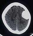
Contrast- enhancing meningioma of the left hemisphere on computed tomography

Large temporal bone meningioma on computed tomography

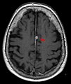
Typical marginal localization of a meningioma on the right (magnetic resonance imaging, T1-weighted with contrast agent)
Falxmeningioma in magnetic resonance imaging: T1 with contrast medium

Meningioma in the spinal canal magnetic resonance imaging. Left T1 sagittal with contrast agent, right T2 coronal


therapy

The neurosurgical removal of the tumor is the treatment of choice. One option here in vascular-rich tumors can be preoperative embolization . Also can possibly radiotherapy or radiosurgery ( Gamma Knife or Cyberknife ) are carried out. If a meningioma cannot be completely removed surgically, the remaining tumor remnants can also be irradiated after the operation in order to reduce the likelihood of tumor growth again. Small meningiomas that do not tend to grow in the elderly do not necessarily need to be removed.
Especially with larger meningiomas with a complex (non-spherical) shape and close relationship to radiation-sensitive organs such as B. The optic nerve or pituitary gland (pituitary gland, controls the hormonal glands in the body), the gentler fractionated stereotactic radiation is recommended. With stereotactic precision irradiation using a linear accelerator, X-rays are generated. A software-controlled micro-multi-leaf collimator (MMLC) adjusts the radiation dose to the shape of the tumor. The multi-leaf collimator consists of movable lead screens, each 3 mm thick, which are individually adjustable and can thus cover and protect healthy tissue. As with gamma knife irradiation, it is not positioned over a metal frame that is screwed onto the head, but rather over a special mask (thermoplastic head mask) that is individually modeled and put on before each treatment session. This form of high-precision radiation is offered in specially equipped departments for radiation therapy. The advantage of splitting the irradiation over several sessions lies in the better possibility of DNA repair of the cells of healthy tissues in the breaks between two irradiation sessions. This recovery (called the fractionation effect) can further reduce the risk of serious side effects from the radiation.
Newer approaches in the treatment of meningiomas deal with the additional use of blood vessel growth or tumor growth inhibiting drugs. Attempts are made to treat more aggressive recurring meningiomas that can no longer be operated on or re-irradiated. However, this approach is still experimental and not a standard procedure.
literature
- Joung H. Lee (Ed.): Meningiomas: Diagnosis, Treatment, and Outcome. Springer, London 2008, ISBN 978-1-84628-526-4 .
- C. Mawrin, A. Perry: Pathological classification and molecular genetics of meningiomas. In: Journal of Neuro-Oncology . Volume 99, Number 3, September 2010, pp. 379-391, ISSN 1573-7373 . doi: 10.1007 / s11060-010-0342-2 . PMID 20809251 . (Review).
- I. Whittle, C. Smith, P. Navoo, D. Collie: Meningiomas. In: The Lancet . 363, 2004, pp. 1535-1543, doi: 10.1016 / S0140-6736 (04) 16153-9 .
- Anne G Osborn: Osborn's Brain: Imaging, pathology and anatomy. Amirsys Publishing, 2013, ISBN 978-1-931884-21-1 .
- M. Kufeld, B. Wowra, A. Muacevic, S. Zausinger, JC Tonn: Radiosurgery of spinal meningiomas and schwannomas. In: Technol Cancer Res Treat. 11 (1), Feb 2012, pp. 27-34.
Web links
- Meningiomas , Deutsche Hirntumorhilfe e. V.
- meningeom.at , Austrian meningioma self-help group, list of treating hospitals and doctors.
Individual evidence
- ↑ LR De Vitis, A. Tedde et al .: Screening for mutations in the neurofibromatosis type 2 (NF2) gene in sporadic meningiomas. In: Hum Genet. 97 (5), May 1996, pp. 632-637.
- ^ DE Reuss, RM Piro et al: Secretory meningiomas are defined by combined KLF4 K409Q and TRAF7 mutations. In: Acta Neuropathol . 125 (3), Mar 2013, pp. 351-358.
- ↑ F. Sahm, J. Bissel et al: AKT1E17K mutations cluster with meningothelial and transitional meningiomas and can be detected by SFRP1 immunohistochemistry. In: Acta Neuropathol. 126 (5), Nov 2013, pp. 757-762.
- ↑ Elizabeth B. Claus, Lisa Calvocoressi et al: Dental x-rays and risk of meningioma. In: Cancer . Volume 118, No. 18, September 15, 2012, pp. 4530-4537, doi: 10.1002 / cncr.26625 .
- ^ AE Elia, HA Shih, JS Loeffler: Stereotactic radiation treatment for benign meningiomas. In: Neurosurgical Focus . 23 (4), 2007, p. E5. PMID 17961042 .
- ↑ Archived copy ( Memento from July 14, 2014 in the Internet Archive )
- ↑ Julia Ahlswede: Aspects of positioning and dose application in stereotactic guided intra- and extracranial radiation therapy. 2005. (edoc.hu-berlin.de)
- ↑ STEREOTACTIC RADIOSURGERY AND RADIOTHERAPY OF MENINGIOMAS . Clinical white paper. PDF version
- ^ Emil Lou, Ashley L. Sumrall et al .: Bevacizumab therapy for adults with recurrent / progressive meningioma: a retrospective series. In: J Neurooncol. 109 (1), Aug 2012, pp. 63-70. PMC 3404217 (free full text); Source of translation