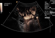
Micturition urosonography
The Miktionsurosonografie is a method of contrast enhanced ultrasound which a radiation-saving alternative to voiding cystourethrogram is used to the vesico-uretero-renal reflux (VUR) , so the backflow of urine from the bladder into the kidneys to check. It is mainly used with children.
Basics
See also main article vesicorenal reflux
The vesico-uretero-renal reflux (VUR) is particularly in children one of the most common causes of febrile urinary tract infections .
A distinction is made in principle:
- Primary VUR: Congenital, mostly anatomical malfunction of the closing mechanism of the ureter opening into the urinary bladder
- Secondary VUR: impaired urine outflow from the bladder, damage / inflammation of the mucous membrane, operations (e.g. after a ureterocele has been slit )
or.
- Low pressure reflux: Reflux even when the bladder is filled
- High pressure reflux: Reflux only when the pressure in the bladder increases during micturition
The reflux can cause germs to get into the kidneys along the urinary tract and thus lead to febrile urinary tract infections . The (primary) VUR in childhood often matures (“matures”) during growth, so that surgical therapy is not necessary immediately after a VUR is detected. Depending on the severity of the VUR, low-dose antibiotic prophylaxis to avoid febrile urinary tract infections and initially a wait-and-see approach is recommended. If check-ups show no more reflux, antibiotic prophylaxis can be discontinued. Persists VUR, a must from the age or constantly appearing febrile urinary tract infection despite antibiotic antireflux (injections of the ureteral orifices , Ureterneueinpflanzung) take place.
Reflux testing
In addition to ultrasound of the urinary tract to document the anatomy and any existing pathological changes, a reflux test must be carried out if febrile urinary tract infections occur, since ultrasound alone cannot reliably detect reflux as the cause.
Micturition Cystourography (MCU)
See also main article voiding cystourethrogram
The MCU is the most common method of testing the VUR. The advantage is that it is easier to assess the urethra in comparison to micturition urosonography. The disadvantage, however, is the radiation dose inevitably associated with an X-ray examination , especially since in this examination the gonads are in the useful beam ( i.e. the full dose of the X-rays is received).
Sonographic reflux examination using MUS
The principle of micturition urosonography (MUS) is the same as that of the MCU : by introducing a contrast medium into the bladder, it can be made visible whether the urine can flow back into the kidneys . The MUS uses special ultrasound contrast media for this purpose (see there: Contrast-enhanced ultrasound ). The contrast medium can be introduced into the bladder by puncturing the urinary bladder or by inserting a urinary catheter through the urethra. Since the catheter is much gentler and less laborious to lay, it has proven to be more practical.
Investigation process
- Ultrasound of the urinary tract to show the anatomy
- Insertion of a urinary catheter under sterile precautions
- If necessary Filling of the urinary bladder with isotonic saline solution at body temperature (0.9%)
- Switching the sonography device to contrast mode
- Instillation of the echo contrast enhancer (e.g. Levovist®, SonoVue®)
- Careful examination of the retrovesical space (to identify first-degree reflux )
- Relocation of the patient, depending on the willingness to cooperate in the prone position, in a sitting position or in the supine position
- Careful examination of both kidneys
- Further filling of the bladder with isotonic saline solution
- Examination while urinating
- If necessary, a second filling, if necessary and if the catheter is still in place
Initial examination

In girls, MUS is recommended as an initial examination and for further follow-up checks. The initial examination of the boy requires the urethra to be shown , as there may be urethral valves there. Only very experienced examiners can do this with MUS, so that the initial examination of boys is often still carried out as a conventional MCU .
literature
- G Zimbaro, G Ascenti, C Visalli, A Bottari, F Zimbaro, N Martino, S. Mazziotti: Contrast-enhanced ultrasonography (voiding urosonography) of vesicoureteral reflux: State of the art. In: Radiol Med (Torino), 2007 Dec, 112 (8), pp. 1211-1224, Epub 2007 Dec 12, PMID 18074195 .
- K. Darge: Voiding urosonography with US contrast agents for the diagnosis of vesicoureteric reflux in children: II. Comparison with radiological examinations. In: Pediatr Radiol. , 2008 Jan, 38 (1), pp. 54-63, Epub 2007 Jul 18, PMID 17639371 .


