urethra
The urethra ( Latin urethra , from Greek ουρήθρα ourḗthrā 'urethra', from ούρα oúra 'urine') is a tubular organ of the urinary and reproductive system of mammals to the exclusion of monotones . It belongs to the urinary tract and begins at the lower end of the urinary bladder located in the pelvis . In male representatives it ends at the tip of the penis on the glans and in female in the vaginal vestibule .
The urethra is primarily the excretion of urine from the bladder, in male mammals in addition, the forwarding of the sperm in the mating , and therefore also here as urinary seed tube is referred to.
The inflammation of the urethra ( urethritis ) is mainly caused by bacteria. In humans, they are mainly transmitted sexually .
function
micturition
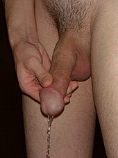
The urethra is used in all mammals with the exception of monotons and in both sexes primarily to drain and excrete the urine, which in mammals collects in the urinary bladder and from there into the urethra ( micturition ). In addition, the closing mechanisms of the urethra, together with immunoglobulins, largely prevent germs from penetrating the interior of the body. The mucous membrane of the urethra in men is only colonized by bacteria in the last third, in women only in the half near the vagina. Mycobacterium smegmatis , corynebacteria , streptococci and Staphylococcus epidermidis belong to this physiological mucosal flora . The sections of the urethra further on the bladder side are sterile.
ejaculation
In male mammals , it also serves to pass on the sperm , which are passed through the spermatic duct ( ductus deferens ) into the urethra and together with secretions from the prostate and the vesicle gland form the sperm . This is transported through the urethra during ejaculation. For this reason, the urethra of male individuals is also known as the urinary-seminal tube .
Sexual stimulation
The urethra is traversed by many nerves and can be viewed as an erogenous zone . Massage or stimulation of the urethra, either manually or by inserting objects, can be experienced as pleasurable for both men and women. Urethral intercourse , in which the male penis is inserted into the dilated female urethra, is very rare .
anatomy
General structure and muscles
|
|
|
|
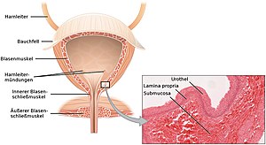
Left : Schematic representation of the anatomical relationships between the urinary bladder and urethra,
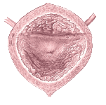
Right : View from the inside of the urinary bladder, the trigonum vesicae and, as the place of origin of the urethra, the central opening of the inner urethral orifice ( ostium urethrae internum ) |
||
Due to its close association with the genital organs, which are differently developed according to gender, the urethra is also different in the sexes. The urethra is a membranous, muscular tube that arises from below the urinary bladder via the inner urethral opening ( ostium urethrae internum ) and from here passes through the pelvic floor in the area of the urogenital diaphragm . Like all urinary pathways , it has a special lining called the urothelium or transitional epithelium . Under the epithelium there is elastic connective tissue and a network of blood vessels ( stratum spongiosum ). Smooth muscles follow further outwards and, on the very outside, connective tissue for embedding in the environment ( adventitia ).
In addition to the smooth muscles, the urethra is surrounded by other muscle parts that enable it to function. Parts of the outer longitudinal muscle layer of the detrusor vesicae muscle , which functions as the expulsion muscle of the urinary bladder, extend from the urinary bladder to the urethra and surround it from the front at the bladder neck together with muscle bundles of the pubovesicalis muscle , which encompass the posterior area of the inner urethral orifice and together the involuntary sphincter of the urinary bladder. This is often seen as the urinary bladder's own sphincter muscle ( sphincter vesicae muscle ). In the transition area from the bladder to the urethra, the lower, thickened angle of the urinary bladder triangle ( Trigonum vesicae ) extends as a muscular uvula vesicae from behind into the inner urethral opening. Fibers of the rectovesicalis muscle attach to the urethra and the urinary bladder from the back.
The striated outer voluntary urethral sphincter ( Musculus urethralis ) surrounds the male urethra with the Musculus sphincter urethrae membranaceae in spiral loops ascending in the area of the Pars membranacea urethrae , in female mammals it arises from the side of the vagina and forms a loop around the urethra. In interaction with the urinary bladder muscles of the urinary bladder triangle, the urinary bladder sphincter and the detrusor vesicae muscle, it makes a significant contribution to urinary retention ( urinary incontinence ) and micturition , whereby the levator ani muscle as pelvic floor can also be involved in urethral occlusion . The urethralis and sphincter vesicae muscles are innervated by the perineal nerves , which arise from the pudendus nerve , while the detrusor vesicae muscle is innervated by the pelvinus nerve.
Between the urethra or its mouth, meatus urethrae , the uretrovaginal septum begins to expand as a periurethral connective tissue space between the female urethra and vagina or vaginal vestibule , vestibulum vaginae . This connective tissue space continues backwards, dorsally into the Halban fascia, also known as the vesicovaginal septum .
Gender-specific anatomy of the urethra
Female mammals

|

|
|
|
Left : Schematic sectional view through the female abdomen with the position of the urethra
Right : Urethral orifice ( Meatus urethrae externus ), including the larger opening of the vaginal entrance ( Introitus vaginae ) |
||


|

|
|
|
Left : Schematic sectional view through the male abdomen with the position of the urethra
Right : Urethral orifice, Meatus urethrae externus , on the glans of the man |
||
The urethra of female mammals runs parallel to the vagina through the pelvic floor and opens into the vulva at the border of the vaginal vestibule and vagina . In women, it is about 2.5 to 4 cm long. In female cloven-hoofed animals there is a blind-ended mucous membrane ( diverticulum suburethrale ) in the area of the mouth , which makes catheterization difficult. The female urethra has a bulge-like or spur-like protuberance ( Carina urethralis vaginae ) on which the actual opening of the urethra, Meatus urethrae externus, is located. Often two rein-shaped, narrow (triangular) tissue folds pull up from this side in the direction of the clitoris; they can become visible during digital examination. The carina urethralis vaginae can, for its part, vary greatly from one individual to the next, protruding to different degrees beyond an imaginary vaginal entrance plane (from a relatively flat profile to a more pronounced one) and resembling a gargoyle . The vaginal entrance, introitus vaginae, is a round tissue edge that lies at the caudal end of the vagina ( carunculae hymenales ) and at the upper end connects to the mouth or the mucous membrane surrounding the urethra, carina urethralis vaginae .
Its initial part near the bladder is lined with urothelium, which merges into multi-layered uncornified squamous epithelium. Occasionally there are mucous glands ( glandulae urethrales ). The mucous membrane forms bays, lacunae urethralis and longitudinal folds that contain the branched, tubular glandulae urethrales . In the distal third, the intraluminal gland ducts of the paraurethral glands, paraurethral glands, can be detected. The closure ( sphincter ) occurs (reflexively) voluntarily by the musculus sphincter urethrae membranaceae , by means of innervation by the nervi perineales from the nervus pudendus .
Periurethral cavernous system
On each side of the actual meatus urethrae externus , the urethral opening, two smaller hump-like hillocks protrude up to the left and right of a channel-like depression or slight notch upwards to the pubic mound. To the side of the bulging carina urethralis vaginae are the paraurethral glands , glandulae paraurethrales ; they have several ducts and open into the end section of the urethra (“intraluminal”) itself as well as laterally (“periurethral”) thereof. They are also called Skene glands (after Alexander Skene ) after their first description or, because of their homology to the male prostate, also prostate feminine .
The glands of the female prostate ( prostate feminina ) or paraurethral gland open into the urethra ; this has several execution routes. Their secretion (see female ejaculation ) is similar in composition and enzyme pattern to the male prostate secretion ( prostate-specific antigen ).
The female prostate is part of a periurethral erectile tissue system, corpus cavernosum urethrae , which also includes the G-spot and, as an intravaginal continuation, the Halban'schen fascia , the Graefenberg zone and the anterior fornix erogenous zone, or AFE zone for short, as additional cavernous tissue . The nerve supply to the female urethra and the surrounding erectile tissue is provided by the vesical plexus (part of the inferior hypogastric plexus ) and the pudendal nerve . Visceral afferents from the urethra run in the splanchnic pelvic nerves.
Male mammals
In all male mammals, the urethra runs through the prostate and penis and opens onto the glans . In many mammals it forms a lobed extension ( processus urethrae ) on the glans , which is very long, especially in the ram . In the stallion, the urethral process sits in a depression, the glans fossa ( fossa glandis ). In men , the urethra ( urethra masculina ) is about 20 to 25 centimeters long and only lined with an urothelium in the initial part. In the middle of the prostate part, in the area of the seminal mound ( Colliculus seminalis ), the urothelium merges into multi-layered, highly prismatic epithelium . Another epithelial transition takes place in the Schiffer's pit ( fossa navicularis urethrae ) to form a multilayered, uncornified squamous epithelium . This epithelium forms a breeding ground for lactic acid bacteria , which create an acidic environment and thus offer protection against infections.
The gender differences in the length of the urethra also have medical consequences: the shorter urethra in female individuals means that the risk of cystitis and urinary incontinence is higher here. Urethral plugs can be used for this in the case of urinary incontinence . In male individuals, the length of the urethra makes it difficult to use a catheter and encourages the retention of urinary stones .
The urethra of the male mammals and man is divided into the following sections:
| Designation in mammals | Name in the man | |
| Pars pelvina (pelvic part) with | Pars praeprostatica (part in front of the prostate ) | Pars praeprostatica (from the urinary bladder wall to the prostate) (1–1.5 cm): It is enclosed by the internal urethral sphincter muscle . |
| Pars prostatica (section of the prostate) | Pars prostatica (section of the prostate) (3–4 cm) | |
| Pars postprostatica with the urethralis muscle | Pars membranacea (pelvic floor part) (1 cm): At this point it is surrounded by the urethralis muscle . The isthmus urethrae is also located here (narrow point in front of the entry into the penis ) | |
| Pars penina ( penis part) | The pars spongiosa is the part in the erectile tissue of the urethra of the penis (approx. 20 cm in men). In animals with a penis bone , the urethra runs in a groove on the abdomen ( sulcus urethrae ) of this bone. Shortly before the mouth, the urethra widens in men ( fossa navicularis urethrae , Schiffer's pit) and then narrows towards the outer mouth ( ostium urethrae externum ) to form a gap. In men, the bulbourethral glands and numerous small mucous glands ( glandulae urethrales ) open here . | |
Evolutive and ontogenetic development
The urethra is an organ that developed together with the urinary bladder exclusively in the Theria , i.e. the marsupial mammals and the higher mammals , while other vertebrates such as birds , reptiles or even the basal mammals, the monotons , have a cloaca .
The urinary bladder and urethra arise in the embryo from the abdominal section of the cloaca, the urogenital sinus . This sinus, in English "cave", has three levels from which the urinary tract in male and female mammals develop. The urinary bladder arises from the upper section, the urethra of female mammals develops from the middle section. In male mammals, this only forms the sections of the urethra that lie in the prostate and pass through the pelvic floor muscles. The section in the penis emerges from the lower level of the urogenital sinus : While this forms the vaginal vestibule in female mammals , it grows with the development of the penis and is enclosed by the erectile tissue of the urethra .
Examination of the urethra
There are various direct and indirect methods of medical examination of the urethra. A simple method, for example to identify injuries or bulges, is scanning ( palpation ). The urethrocystoscopy is an endoscopic procedure in which the endoscope is inserted into the urethra and slowly advanced into the bladder. This enables the precise localization of constrictions, bulges or tumors. The procedure is used to establish indications for all interventions in the area of the bladder neck, for example for transurethral resection of the prostate.
Imaging methods include (retrograde) urethrography , in which the urethra is filled with an X-ray contrast medium to identify urinary stones, foreign bodies, constrictions, malformations, tumors and other abnormalities, as well as the micturition cystourethrogram , in which the bladder is passed through a catheter with contrast medium is filled. During the excretion of the contrast agent, as in urethrography, bulges or obstructions to the drainage of the urethra can be recognized. For the ultrasound examination , the urethra must first be filled with water or lubricant. This method is used to clarify urethral strictures.
Indirect examinations for the detection of infectious agents can be carried out via a urethral swab, i.e. the removal of a swab sample, or the examination of the urethral discharge. The examination of the urine is not very meaningful because the germ content of the urethral secretion is reduced too much by the sterile flowing urine.
Malformations, diseases and injuries
Malformations, diseases and injuries can affect the urethra of all mammals, whereby the infections in particular are different and different, sometimes species-specific, pathogens are possible triggers. The following representations are primarily related to humans, but can be applied to other mammals accordingly.
Malformations

As with other organs, the lower urinary tract and directly the urethra can be affected by malformations that develop during early childhood development due to various factors. All malformations of the urethra are treated and corrected surgically if they cause symptoms.
If the urethra is not created, it is called an agenesis ; if it is blocked at one point, it is called an atresia . Both malformations are rare and only compatible with life if another possibility of urine drainage remains, such as an open urachus . In some neonates, usually male, urethral valves remain , which impede the flow of urine.
One of the most common malformations of the male genital organs is hypospadias . In this case, the urethra is incompletely closed lengthways and opens on the underside (ventral side) of the penis. It also occurs very rarely in girls, in which case the urethra opens into the vagina . The dorsal gap, epispadias , is also very rare. In boys, the gap is on the top of the penis; in girls, the clitoris is usually split. Epispadias usually occur with bladder exstrophy .
Also, a doubling of the urethra or the formation of a rudimentary urethra as an attached (accessory) ductus paraurethralis occur.
Diverticulum, fistula and prolapse
As a urethral diverticulum bulges are called, which can occur in all sections usually at the bottom of the urethra. These can be congenital or later develop in response to an inflammation-related urethral stricture prior to that stricture. Diverticula are usually identified by chronic urethritis with dysuria as well as hematuria or urinary incontinence .
Fistulas are connections between a hollow organ and another organ. They too can be congenital or acquired. In women, acquired urethrovaginal fistulas , i.e. fistulas between the urethra and vagina , are usually the result of abscesses , for example from inflammation of the urethral diverticula, or injuries from accidents and operations.
In the case of a urethral prolapse ( urethral prolapse), the urethral mucous membrane protrudes from the outer urethral orifice. Urethral rolapse occurs primarily in young girls; in animals, males of short-headed dog breeds are particularly affected. The cause is not clear, treatment is carried out by surgically removing the prolapsed mucous membrane.
Inflammation and infection
The most common disease of the urethra is inflammation of the urethral mucosa, urethritis , which may also affect deeper layers and is then referred to as periurethritis and cavernitis . The inflammations are divided into infectious, mechanical and allergic urethritis according to the cause. A fourth group is the urethritis in general diseases.
Infectious urethritis is the largest group. They can occur as part of a general urinary tract infection or they can be caused by specific pathogens. Here, gonorrheic urethritis ("gonorrhea") caused by Neisseria gonorrhoeae , is differentiated from non-gonorrhoeic urethritis , behind which all other pathogens stand. In this group, bacterial infections are leading, especially those caused by chlamydia ( Chlamydia trachomatis ). Non-gonorrheic urethritis is the most common sexually transmitted disease in developed countries. The postgonorrhoische urethritis is a mixed infection of Neisseria gonorrhoeae and Chlamydia trachomatis . Less common are viruses or fungi trigger inflammation, but these pathogens are more likely in individuals with weakened immune systems appear. The Trichomonas vaginalis parasite colonizes the vagina and urethra. The trichomoniasis it causes is also a sexually transmitted disease. After the pathogen has been determined, infectious urethritis is treated with the help of anti-infectives such as antibiotics , antivirals or antimycotics against the specific group of pathogens.
Mechanical urethritis is the result of mechanical irritation, for example from the insertion of a urinary catheter . Catheter urethritis is then often associated with a bacterial infection. Allergic urethritides are primarily intolerance reactions to medication, as well as food and beverages . These can occur in men whose sexual partner vaginally applied contraception use.
In addition to the causes mentioned, inflammation of the urethral mucous membrane can also occur as a result of other acute and chronic general diseases such as diabetes mellitus , Reiter's disease or typhus abdominalis .
Tumors
Polyps are benign tumors of the mucous membrane that usually grow on peduncles. They mostly occur in the urethra in postmenopausal women and impede the flow of urine. They need to be demarcated from urethral caruncles. These are mucous membrane-coated, easily bleeding tumors of the connective tissue interspersed with inflammatory cells, which mostly form near the urethral orifice and can emerge from it (prolapse). They are also common in older women.
The transitional cell carcinoma (urethral cancer), however, is a rare malignant tumor of the urethra, usually called squamous occurs urethral mucosa. Women are affected three times as often as men, with the frequency peak between the ages of 50 and 65. The symptoms are mostly unspecific; blood or bloody fluid can leak from the urethra. If the carcinoma is located close to the bladder, perineal pain , abscesses, and fistulas can also occur. In women, diverticula, caruncle and chronic inflammation seem to promote the development of urethral cancer, in men the disease can be a long-term consequence of infections and injuries.
Injuries and foreign bodies
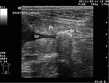
Injuries to the urethra are divided into "posterior" and "anterior" urethral injuries, which differ in terms of their cause. By posterior urethral injuries are meant injuries to the urethra above its passage through the pelvic floor. They mostly arise when pelvic fractures are caused by massive violence, i.e. in traffic accidents and falls from great heights. It affects the urethra of men more often than the urethra of women. Anterior urethral injuries are injuries to the urethra below the pelvic floor. In adults, medical measures are the leading causes, i.e. the insertion of instruments and the insertion of urinary catheters and lying for too long. Here, too, male urethras are more frequently affected, especially catheters that are left in place for too long can lead to strictures of the penile part of the urethra. Injuries from other inserted objects, such as sexual stimulation, are rare and affect the urethra close to its opening. Traumatic anterior urethral injuries are more common in children than in adults, primarily as a result of impact trauma to the perineum . In men who suffer a penile rupture , the urethra is also affected in 20% of cases.
Also urinary stones ( urolithiasis ) that are swept from the bladder into the urethra or only occur in the urethra come before and can lead to a narrowing or stricture lead. These are pathological solid structures ( concretions ) that can form in the urine from mineral salts.
The Vandelliinae ("urethral catfish") that occur in Brazil can penetrate the urethra, get stuck there and die, leading to an obstruction of the urethra.
Piercings and other modifications
As with other parts of the genital region, the human urethra can also be included in the intimate jewelry in the form of various piercings , which are then realized as jewelry with a ball closure ring , a segment ring and a curved barbell . The most famous form of these genital piercings is the man's Prince Albert piercing , which runs from the urethra through the lower penis wall. Modifications are the Reverse Prince Albert Piercing with an exit at the top of the penis and the Dolphin Piercing with two openings in the lower urethra. The Ampallang is a piercing that is pierced horizontally through the glans and usually crosses the urethra. The apadravya, on the other hand, is pricked vertically and crosses the urethra, provided it has been placed in the middle, which is the case with most apadravyas. Another piercing jewelry made by men is the Prince's Wand , which is inserted into the urethra as a pin and is usually attached to the glans (pinless) with a Prince Albert piercing, an Ampallang or an Apadravya or a ring. The female equivalent of the Prince Albert piercing is the Princess Albertina piercing , which runs from the urethral opening to the vaginal opening.
In the medical literature there are isolated references to injuries or complications that can be caused by piercings with a urethral passage. In addition to inflammation and allergic reactions, these include tears, fistulas or other injuries.
In addition to piercings, there are other modifications that mainly affect the penis and the male urethra. A urethral stretch for sexual stimulation can be performed with the help of dilators . The urethra can be stimulated using a urethral vibrator or urethral plug . In the case of bifurcation , the penis is divided to varying degrees from the glans, and the urethra is also affected. In the case of the subincision , this division takes place only on the underside through the split of the urethra and the underside of the penis, but this can extend to the penis attachment.
Intimate jewelry and modifications that include the urethra are essentially used as jewelry for the aesthetic change in the genital area. In addition, they can have a function of increasing stimulation during sexual intercourse and masturbation .
literature
- Pschyrembel Medical Dictionary. 266th edition, de Gruyter, Berlin 2014, ISBN 978-3-11-033997-0 .
- Helga Fritsch, Wolfgang Kühnel: Pocket Atlas Anatomy. Volume 2 . 9th, revised edition. Thieme, Stuttgart 2005, ISBN 3-13-492109-X , p. 262 .
- Uwe Gille, Franz-Viktor Salomon: urethra, urethra . In: Franz-Viktor Salomon u. a. (Ed.): Anatomy for veterinary medicine . Enke, Stuttgart 2015, ISBN 978-3-8304-1288-5 , pp. 391 .
- Theodor H. Schiebler (Ed.): Anatomie. Histology, history of development, macroscopic and microscopic anatomy, topography . 9th edition. Springer, Berlin / Heidelberg 2005, ISBN 3-540-21966-8 , pp. 611-613 .
- Richard Hautmann (Ed.), Jürgen E. Gschwend: Urology. 5th edition. Springer-Verlag , Berlin / Heidelberg 2014, ISBN 978-3-642-34318-6 .
- Jürgen Sökeland, Herbert Rübben u. a .: Pocket textbook urology. 14th, completely revised edition, Thieme, Stuttgart a. a. 2008, ISBN 978-3-13-300614-9 .
supporting documents
- ↑ Michael Wilson: Microbial Inhabitants of Humans: Their Ecology and Role in Health and Disease . Cambridge University Press, 2005, ISBN 978-0-521-84158-0 , pp. 195 .
- ^ Nicholas J. Vardaxis: Immunology for the Health Sciences . Macmillan Education AU, 1996, ISBN 0-7329-3092-8 , pp. 15 .
- ↑ "How can I stimulate my urethra?" - look
- ↑ The G-spot was yesterday! Now the urethra is stimulated - Viva
- ↑ U-Spot: Hotter than the G-Spot? - Fem
- ↑ a b c d W. Kahle, H. Leinhardt, W. Platzer (eds.): Pocket Atlas of Anatomy for Study and Practice. Volume 2: Internal Organs. 5th edition, Thieme, Stuttgart 1986, ISBN 3-13-492105-7 , pp. 268-269.
- ↑ W. Kahle, H. Leinhardt, W. Platzer (ed.): Pocket Atlas of Anatomy for study and practice. Volume 2, Stuttgart 1986, pp. 266-267.
- ↑ Angelika Strunk: Fascial Osteopathy: Basics and Techniques. Georg Thieme Verlag, Stuttgart 2015, ISBN 978-3-8304-7922-2 , p. 74
- ↑ Beate Carrière: pelvic floor. Georg Thieme Verlag, Stuttgart 2012, ISBN 978-3-13-170332-3 , p. 536
- ^ JW Huffman: The detailed anatomy of the paraurethral ducts in the adult human female. Am J Obstet Gynecol, 1948; 55: 86-101
- ↑ a b Renate Lüllmann-Rauch: Pocket textbook histology . 4th edition. Georg Thieme Verlag, Stuttgart 2012, p. 488. ISBN 3-13-129244-X
- ↑ Thomas Deller: Histology - The textbook Elsevier Health Sciences, Munich 2018, ISBN 978-3-4371-8366-9 , p. 491 [1] books.google.de
- ↑ Florian Wimpissinger: The female prostate - fact or myth? In: urology. Volume 2/07, p. 19.
- ↑ Per Olov Lundberg: The peripheral innervation of the female genital organs. Sexology 9 (3) 2002, 98-106 [2]
- ↑ Werner Böcker et al .: Pathology , 5th edition. Urban & Fischer, Munich 2012, ISBN 978-3-437-42384-0 , p. 722.
- ↑ Renate Lüllmann-Rauch: Pocket textbook histology . 4th edition. Georg Thieme Verlag, Stuttgart 2012, p. 506.
- ^ A b Nadja Møbjerg: organs of osmoregulation and excretion in: W. Westheide and R. Rieger: special zoology. Part 2: vertebrates or skulls. Spektrum Akademischer Verlag, Munich 2004, ISBN 3-8274-0307-3 , p. 151.
- ↑ Thomas W. Sadler: Medical Embryology . Translated from the English by Ulrich Drews. 11th edition, Thieme, Stuttgart 2008, ISBN 978-3-13-446611-9 , p. 265, p. 319 f.
- ↑ Jürgen Sökeland, Herbert Rübben: Pocket textbook urology. 14th edition, Stuttgart a. a. 2008, p. 46. ISBN 3-13-300614-2
- ↑ Jürgen Sökeland, Herbert Rübben: Pocket textbook urology. 14th edition, Stuttgart a. a. 2008, p. 85.
- ↑ Jürgen Sökeland, Herbert Rübben: Pocket textbook urology. 14th edition, Stuttgart a. a. 2008, p. 72.
- ↑ Richard Hautmann (Ed.), Jürgen E. Gschwend: Urology . Berlin / Heidelberg 2014, p. 58.
- ↑ Richard Hautmann (Ed.), Jürgen E. Gschwend: Urology . Berlin / Heidelberg 2014, p. 45.
- ^ Frank Hegenscheid: Urethradiagnostik . In: Ralf Tunn, Engelbert Hanzal, Daniele Perucchini (eds.): Urogynaecology in practice and clinic . Walter de Gruyter, 2009, ISBN 978-3-11-020688-3 , p. 93-94 .
- ↑ a b c Keyword "Urethral malformations" In: Pschyrembel Medical Dictionary. 266th edition, Berlin 2014, p. 854.
- ↑ Jürgen Sökeland, Herbert Rübben: Pocket textbook urology. 14th edition, Stuttgart a. a. 2008, p. 184.
- ↑ Jürgen Sökeland, Herbert Rübben: Pocket textbook urology. 14th edition, Stuttgart a. a. 2008, p. 182.
- ↑ Jürgen Sökeland, Herbert Rübben: Pocket textbook urology. 14th edition, Stuttgart a. a. 2008, p. 179.
- ↑ Jürgen Sökeland, Herbert Rübben: Pocket textbook urology. 14th edition, Stuttgart a. a. 2008, p. 180.
- ↑ Jürgen Sökeland, Herbert Rübben: Pocket textbook urology. 14th edition, Stuttgart a. a. 2008, p. 178 f.
- ↑ a b Keyword “urethral diverticulum” In: Pschyrembel Medical Dictionary. 266th edition, Berlin 2014, p. 854.
- ↑ Keyword “urogenital fistula” In: Pschyrembel Medical Dictionary. 266th edition, Berlin 2014, p. 2205.
- ↑ HU Braedel et al .: Traumatology of the urogenital tract . Springer, 2013, ISBN 978-3-642-80573-8 , p. 300.
- ↑ Christina Scheibel: Surgical correction of a urethral prolapse in a French bulldog. In: Kleintierpraxis Volume 62, 2007, Issue 1, pp. 15-19.
- ↑ Keyword "Urethritis" In: Pschyrembel Medical Dictionary. 266th edition, Berlin 2014, p. 2203.
- ↑ a b Richard Hautmann (Ed.), Jürgen E. Gschwend: Urologie . Heidelberg 2014, p. 149.
- ↑ Herbert Hof, Gernot Geginat: Infections of the kidney and the lower urinary tract . In: Herbert Hof, Rüdiger Dörries (ed.): Medical microbiology . 5th edition. Thieme, Stuttgart 2014, ISBN 978-3-13-125315-6 , p. 649.
- ^ Hans U. Schmelz, Christoph Sparwasser, Wolfgang Weidner: Specialist knowledge of urology . 1st edition, Springer-Verlag, Heidelberg 2006, ISBN 978-3-540-20009-3 , p. 63.
- ↑ a b Keyword "urethritis, not gonorrheic" In: Pschyrembel Medical Dictionary. 266th edition, Berlin 2014, p. 2203.
- ↑ Keyword "urethritis, postgonorrheic" In: Pschyrembel Medical Dictionary. 266th edition, Berlin 2014, p. 2203.
- ^ Otto Braun-Falco, Gerd Plewig, Helmut Heinrich Wolff, Walter Burgdorf, Michael Landthaler: Dermatology and Venereology . 5th edition. Springer, Berlin 2005, ISBN 978-3-540-26624-2 , pp. 288 .
- ↑ Keyword "Urethral polyp" In: Pschyrembel Medical Dictionary. 266th edition, Berlin 2014, p. 854.
- ↑ Keyword “Urethral Caruncle” In: Pschyrembel Medical Dictionary. 266th edition, Berlin 2014, p. 854.
- ^ A b Hans U. Schmelz, Christoph Sparwasser, Wolfgang Weidner: Specialist knowledge of urology . Heidelberg 2006, p. 210.
- ^ Hans U. Schmelz, Christoph Sparwasser, Wolfgang Weidner: Specialist knowledge of urology . Heidelberg 2006, p. 212.
- ↑ Richard Hautmann (Ed.), Jürgen E. Gschwend: Urology . Berlin / Heidelberg 2014, p. 297.
- ↑ JL Breault: Candiru: Amazonian parasitic catfish . In: Journal of Wilderness Medicine , 2, 1991, No. 4, pp. 304-312.
- ↑ a b c d e f Michael Waugh: Body piercing: where and how. In: Clinics in Dermatology , 25 (4), July / August 2007; Pp. 407-411. doi: 10.1016 / j.clindermatol.2007.05.018
- ↑ Martin Kaatz, Peter Elsner, Andrea Bauer: Body-modifying concepts and dermatologic problems: tattooing and piercing. In: Clinics in Dermatology 26 (1), January / February 2008; Pp. 35-44. doi: 10.1016 / j.clindermatol.2007.10.004


