Urinalysis
The urinalysis or urogenostics is one of the oldest methods of examining the presence, severity and course of diseases of the kidneys and urinary tract .
| Classification according to ICD-10 | |
|---|---|
| R82.- | Other abnormal urine findings |
| R82.0 | Chyluria |
| R82.1 | Myoglobinuria |
| R82.2 | Bilirubinuria |
| R82.3 | Hemoglobinuria |
| R82.4 | Acetonuria |
| R82.5 | Elevated urine levels for drugs, pharmaceuticals, and biologically active substances |
| R82.6 | Abnormal urine values for substances primarily of non-medical origin |
| R82.7 | Abnormal results on microbiological urine examination |
| R82.8 | Abnormal findings on urine cytological and histological examination |
| R82.9 | Other and unspecified abnormal urine findings |
| R81 | Glucosuria |
| R80 | Isolated proteinuria |
| ICD-10 online (WHO version 2019) | |
In antiquity and the Middle Ages until well into the early modern times and in some cases into the 19th century, it was carried out as uroscopy or urine inspection (an examination and odor test of the spontaneously emptied urine ) for diagnostic purposes. In doing so, reference was mainly made to humoral pathology , the doctrine of humors according to Hippocrates of Kos (approx. 460 to approx. 370 BC) and Galen of Pergamon (approx. 129 to approx. 216 AD). Even today in Unani medicine , urination is still used with the naked eye.
Starting at the beginning of the 19th century, scientific urine testing was finally established in the 20th century.
Today, in most cases, a urine test strip is used first, which enables a quick, simple and inexpensive analysis of the urine for the presence of red blood cells (erythrocytes) , white blood cells (leukocytes) , protein , nitrite , glucose and other substances.
Test strips
If the test strip shows abnormal results, especially if red or white blood cells are detected, the urine is centrifuged and the urine sediment is examined under the microscope .
Red blood cells in the urine indicate bleeding from the kidneys and urinary tract and can occur in cases of kidney cancer , urinary stones, or diseases of the kidney corpuscle (usually glomerulonephritis ). In about a third of the cases, however, no cause can be found even with careful examination .
White blood cells in the urine usually indicate a urinary tract infection , especially if there is pain when urinating and nitrite is detectable on the test strip.
The most common causes of protein in urine test strips are diseases of the kidney corpuscle such as diabetic nephropathy , nephrosclerosis or glomerulonephritis. For further diagnostics, the protein excretion is quantified using chemical methods , and the different proteins are characterized by electrophoresis .
A large number of other determination methods exist for special questions.
Urine sample
Severe physical exertion (long-distance running, soccer game) should be avoided 72 hours before the urine sample is given. A urine test should not be performed during menstruation . Women should use a tampon for discharge. The opening of the urethra should be washed off. The first portion of urine is discarded. In order to reduce the accumulation of cells and secretions from the urethra and vagina , so-called midstream urine is used for the analysis.
Particles in the urine dissolve quickly, especially if the urine is alkaline or dilute (low specific gravity , low osmolality ). Ideally, the urine sample should be examined within 2 to 4 hours. If this is not possible, the urine can be stored at temperatures between +2 ° C and +8 ° C; however, this favors the precipitation of urate and phosphate crystals. Alternatively, the urine can be preserved by adding formaldehyde or glutaraldehyde , but this process of fixation can lead to changes in urine components.
Physical Properties
colour
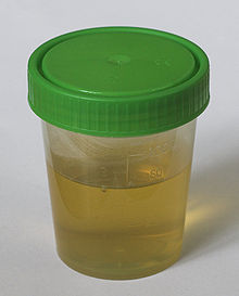
The normal color of urine ranges from light yellow to dark yellow, or it is amber in color.
Illnesses, drugs and food can cause the urine to change color:
- Diseases
-
- Admixtures of blood ( macrohematuria ), hemoglobin ( hemoglobinuria ) and myoglobin ( myoglobinuria ) can discolour the urine pink, red, brown or black.
- Jaundice (icterus) turns yellow or brown in color.
- A massive excretion of uric acid crystals ( crystalluria ) turns the urine pink.
- In porphyria and alkaptonuria , the urine is red and turns black when left standing.
- With metastatic malignant melanoma a black discoloration of the urine can occur in about 15% of cases. The cause is the increased excretion of 5: 6 hydroxy indole , an intermediate product in the formation of melanin from tyrosine (melanuria, Fig. From)
- Medication
-
- Rifampicin turns yellow-orange to red in color.
- Phenytoin turns the urine red.
- Aminophenazone and phenolphthalein can make the urine red
- Chloroquine and nitrofurantoin color the urine brown.
- Triamterene and blue food colors, as well as propofol (with certain genetic variations) can lead to a green color, as well as indomethacin , amitriptyline , listerine, flutamide , and an infection with Pseudomonas aeruginosa
- Metronidazole , methyldopa , imipenem cause the urine to darken when standing.
- food
-
- Beetroot and blackberries can discolour urine red.
- Senna and rhubarb can turn yellow to brown and red.
Cloudiness
Urine is usually clear. Turbidity can be caused by a variety of different particles. Most of these are erythrocytes , leukocytes , bacteria , squamous cells , lipids or crystals. Secretions from the genital area often cause opacity. In urogenital tuberculosis , cheesy material can cloud the urine.
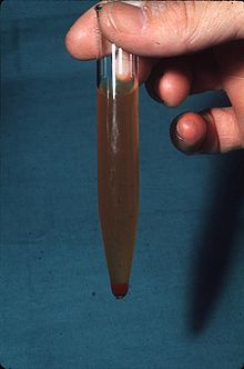
The admixture of chylus (fatty lymph fluid ) with the urine leads to a white cloudiness, especially after high-fat meals ( chyluria ). Chyluria occurs when there is a pathological connection between the lymphatic and genitourinary systems . Causes are filariasis , urogenital tuberculosis, schistosomiasis , injuries , pregnancy , congenital malformations , aortic aneurysms , surgical interventions and inflammation of the mesenteric lymph nodes . (Fig. Below)
odor
A pungent odor in the urine indicates an infection with bacteria that produce ammonia .
Some rare diseases cause a characteristic odor in the urine.
- Maple syrup disease results in a maple syrup odor in urine.
- Phenylketonuria causes a musty or mouse-like odor of urine.
- Isovaleric acidosis causes urine to smell sweat.
- Hypermethioninemia leads to a smell of rancid butter or fish.
- Ketones have a sweet or fruity smell.
The consumption of vegetable asparagus leads to a special smell, for which an enzyme is responsible that breaks down the aromatic substance aspartic acid (1,2-dithiolane-4-carboxylic acid) in the urine . During this process, sulfur-containing compounds are released, which are then excreted.
relativ density
The relative density of urine can be determined using several methods:
specific weight
The specific gravity of the urine depends on the amount of substances dissolved in the urine. The determination is carried out using a urinometer, a countersunk spindle with a scale between 1,000 and 1,060, or a refractometer . The urinometer is quick and easy, but apart from peri-operative, anesthesiological situations (neurosurgical operations) it is rarely used.
Osmolarity
The osmolarity of urine depends on the number of dissolved particles. The measurement is made by means of an osmometer , e.g. B. by determining the lowering of the freezing point.
If more osmotically active particles are filtered into the primary urine , there is an increase in urine osmolarity and urine volume ( osmotic diuresis ). Examples:
- In diabetes mellitus , an increased glucose concentration in the urine can lead to osmotic diuresis.
- Mannitol also leads to osmotic diuresis and can therefore be used as a diuretic .
If the kidney can no longer concentrate the urine sufficiently due to advanced kidney disease, the osmolarity in the urine is the same as in the plasma ( isosthenuria ).
If more water is excreted in the urine, e.g. B. after increased fluid intake ( polydipsia ) or due to diabetes insipidus , the osmolarity in the urine decreases ( water diuresis ).
Refractometry
With the aid of a refractometer capable of refractive index of urine can be determined. This is a measure of the osmolarity of the urine. The test is easy to perform and only requires a drop of urine.
Dry chemistry
An approximate determination of the osmolality is also possible using urine test strips .
In the underlying reaction , a complexing agent releases protons in the presence of cations , which lead to a color change in the indicator bromothymol blue .
If the urine pH is above 6.5, the osmolality is underestimated; if the protein concentration exceeds 7 g / l, the osmolality is overestimated. The underlying reaction only records ions , but not important osmotically active non-ionized molecules such as glucose or urea . For these reasons, there is poor agreement with other methods of determining osmolality.
Chemical properties
pH
The urine test strip is usually used to determine the pH of the urine. The indicator covers a pH range between 5 and 9. If the urine pH exceeds or falls below this range or if a more precise determination of the pH value is required, the measurement must be carried out using a pH meter .
hemoglobin
→ Main article: hematuria
Blood in the urine ( hematuria ) is detected with the urine test strip through the red blood pigment ( hemoglobin ). The detection reaction uses the peroxidase activity of the heme group, which catalyzes the reaction between peroxide and a dye. In the presence of erythrocytes , green spots form; in the presence of free hemoglobin, a homogeneous green color change occurs.
False positive results occur with hemolysis with hemoglobinuria , rhabdomyolysis with myoglobulinuria and with high concentrations of bacteria with peroxidase activity such as enterobacteria , staphylococci and streptococci .
False negative results can occur in the presence of reducing substances. Thus, in the presence of ascorbic acid , e.g. For example, if you take large amounts of vitamin C, you will miss a mild hematuria.
The sensitivity of the test strip for the detection of hemoglobin is 95–100%, the specificity 65–93%.
Glucose
→ Main article: Glucosuria
In the urine test strip, glucose is first oxidized to glucuronic acid and hydrogen peroxide . In a second step, which is catalyzed by a peroxidase , hydrogen peroxide reacts with a color reagent. The test strip allows semi-quantitative detection. If an exact determination of the glucose concentration is required, enzymatic determination methods are used.
Only when the blood sugar is higher than the kidney threshold does glucose pass into the urine and can be detected there. If the glucose concentration in the urine is more than 15 mg / dl (0.8 mmol / l), one speaks of glucosuria . The causes of glucosuria are increased blood sugar levels ( diabetes mellitus ) or reduced reabsorption of glucose from the primary urine in diseases of the kidney tubules ( diabetes renalis ).
False negative results are obtained in the presence of ascorbic acid and bacteria , false positive results can be caused by oxidizing cleaning agents and hydrochloric acid .
protein
→ Main article: Proteinuria
If the excretion of protein in the urine ( proteinuria ) is more than 150 mg / 24 h for a period of more than three months, chronic kidney disease is present.
- The most common causes of proteinuria over 1000 mg / 24 h are diabetic nephropathy , glomerulonephritis and chronic transplant nephropathy .
- If the proteinuria is above 3000 mg / d and there are edema and hyperlipidemia , one speaks of a nephrotic syndrome .
- In nephrosclerosis , interstitial nephritis , cystic kidneys , transplant rejection and drug-induced kidney damage, proteinuria is usually less than 1000 mg / 24 h or absent entirely.
The level of proteinuria correlates with the rate at which renal function is lost. A decrease in proteinuria with therapy indicates a response to treatment.
There are three ways to detect proteinuria:
Protein detection using test strips
The detection reaction is based on the fact that proteins in a buffer system lead to a change in the pH value that is proportional to the concentration of the protein. The pH change was made visible by a pH-dependent color change. This detection method has a high sensitivity to albumin , but only a very low sensitivity to other relevant proteins such as tubular proteins or free light chains .
The test strip only allows a semi-quantitative determination of the protein concentration, which is indicated on a scale from 0 to +++.
An albumin excretion of less than 300 mg / 24 h or less than 200 mg / l, which is known as microalbuminuria, which is particularly relevant in diabetics , cannot be detected by the test strips usually used.
24 hour protein excretion
The urine is collected over 24 hours. At the beginning of the collection period, the urinary bladder must be completely emptied into the toilet, from this point the urine is completely collected in a collection vessel, exactly 24 hours after the start of the collection period, the bladder must be completely emptied into the collection vessel. The concentration of total proteins in the urine can be determined by the biuret reaction , turbidimetry or nephelometry . The protein excretion is given in mg (or g) per 24 hours.
The protein determination in the 24-hour urine collection is the reference method for the protein determination in urine. However, because of the relatively complex collection rules, errors often occur when collecting the urine precisely.
Bacteria can multiply during the collection period. In addition, cellular components of the urine break down during this period. The urine collection must therefore not be used for examining the urine sediment or for microbiological diagnostics.
Protein / creatinine quotient in spontaneous urine
In order to avoid the difficulties of determining the protein excretion over 24 hours, the protein concentration in spontaneous urine can also be related to the creatinine concentration in the urine sample. The protein concentration is then given in mg (protein) / mg (creatinine) or mg (protein) / g (creatinine). The normal value is below 0.07 mg / mg.
There is a good correlation between the protein / creatinine quotient and the 24 hour protein excretion. However, the correlation may be less accurate for protein concentrations above 1 g / l. To date, there have been no studies of the role of the protein / creatinine ratio in monitoring the treatment of diseases associated with proteinuria. In cats, the quotient is an important criterion for assessing chronic kidney failure .
SDS polyacrylamide gel gradient electrophoresis (SDS-PAGE)
→ Main article: SDS-PAGE
Sodium lauryl sulfate (SDS) is added to the urine . This denatures the urinary proteins and can be separated according to their molar mass by electrophoresis on a polyacrylamide gel .
The SDS-PAGE records all urinary proteins and enables the distinction between glomerular proteinuria, tubular proteinuria and prerenal proteinuria. SDS-PAGE does not allow quantification of proteinuria and must therefore always be combined with a quantitative protein determination (24 h protein excretion or protein / creatinine quotient).
- Tubular proteinuria
- In the kidney corpuscles , small-molecule proteins ( α 1 -microglobulin , β 2 -microglobulin , retinol-binding protein , β-NAG ) are filtered into the primary urine and then reabsorbed through the proximal tubular cells of the proximal tubule (renal tubules) . In the case of diseases of the renal tubule system, reabsorption is reduced, and small-molecule proteins can then be detected in the urine.
- Tubular proteinuria indicates interstitial nephritis , pyelonephritis , transplant rejection , acute kidney failure or hereditary diseases of the tubular system such as: B. De Toni Fanconi syndrome .
- Glomerular proteinuria
- If higher molecular proteins appear in the urine, this indicates a defect in the basement membrane of the kidney corpuscle. In the early stages of the disease, proteins with an average molecular weight range of 50–70 kDa ( albumin , transferrin ) can be detected in the urine (selective glomerular proteinuria). In advanced disease, high molecular weight proteins such as B. Immunoglobulin G (unselective glomerular proteinuria).
- Selective glomerular proteinuria suggests minimal change glomerulonephritis , focal sclerosing glomerulonephritis, and early perimembranous glomerulonephritis .
- An unselective glomerular proteinuria with protein excretion> 3000 mg / 24 h suggests proliferative glomerulonephritis , diabetic nephropathy or amyloidosis . Protein excretion <300 mg / 24 h indicates a residual condition after glomerulonephritis , protein excretion <120 mg / day is not evidence of kidney disease.
- Prerenal proteinuria
- In monoclonal gammopathies , large amounts of free light chains can be produced. The free light chains are filtered in the glomerulus and reabsorbed in the proximal tubule. If the filtered amount of free light chains exceeds the capacity of the tubular system for reabsorption, the free light chains appear in the urine ( Bence-Jones proteinuria ).
- Mixed proteinuria
- In advanced kidney disease, both kidney corpuscles and tubules are affected. One then finds mixed forms between glomerular and tubular proteinuria: advanced glomerulonephritis, diabetic nephropathy, nephrosclerosis and amyloidosis.
Urine proteome
→ Main article: Proteomics
The analysis of the urine proteome is an experimental method with which the entirety of the proteins present in the urine is examined. To do this, the proteins are separated using different methods, then ionized and analyzed using mass spectrometry . Two-dimensional gel electrophoresis , liquid chromatography , selective protein adsorption, capillary electrophoresis and protein arrays are used as separation processes . Characteristic protein patterns were observed in IgA nephropathy , vasculitis, and diabetic nephropathy .
Leukocyte esterase
In urine leukocytes existing set indoxyl - esterases free if they burst. This esterase activity can be detected using urine test strips. Cells burst particularly easily in alkaline urine or urine of low density, which is why the test strip is often positive, whereas microscopic examination cannot detect any leukocytes. In contrast, a high density of urine prevents the lysis of leukocytes and thus reduces the sensitivity of the esterase test strip. False negative results can also occur with a high concentration of glucose or protein and in the presence of antibiotics ( cephalotin , tetracycline , cefalexin , tobramycin ). False positive results are rare and occur e.g. B. in the presence of formaldehyde . The sensitivity of the test is 76–94%, the specificity 68–81%.
nitrite
Nitrite is detected using urine test strips and gives an indication of a bacterial urinary tract infection . Most gram-negative bacteria that can cause urinary tract diseases have nitrate reductases , which they can use to reduce nitrates to nitrites . However, there are important pathogens causing urinary tract infections that have little or no activity of nitrate reductase, such as Pseudomonas , Staphylococcus epidermidis and enterococci . In addition, the test can only respond if a sufficient amount of nitrates is ingested with food (e.g. through vegetables) and the urine remains in the urinary bladder for a sufficiently long time.
The sensitivity of the test is therefore low, while the specificity is good at> 90%.
Bile pigments
Urine test strips can also be used to detect urobilinogen and bilirubin in liver disease . In practice, however, this method is no longer relevant, since liver enzymes and bilirubin are determined in the blood when the liver and biliary tract are diseased .
Ketones
→ Main article: Ketonuria
Ketones can be detected using urine test strips through a reaction between nitroprusside with acetoacetic acid and acetone . Ketones in the urine indicate ketosis or ketoacidosis in diabetes mellitus , hunger , vomiting or strenuous physical activity.
microscopy


→ Main article: Urine sediment
The microscopic examination of the urine sediment is an indispensable part of the urine examination and supplements the physical and chemical urine examination with indispensable information.
Methods
The patient is instructed to completely empty the bladder first thing in the morning. In the night urine, the cells present in the urine can dissolve during the long period in the bladder. Therefore, the second morning urine in a disposable collection is collected for the study urine sediment after previously the first milliliter of urine stream were discarded to disturbing admixtures from the urethra to remove ( midstream urine ). The urine sample is then examined within 2-3 hours. For this purpose, 10 ml of the urine are centrifuged for 10 minutes at a speed of 2000 rpm; the supernatant is discarded, the sediment is resuspended and examined with a phase contrast microscope . Crystals and fat droplets can be identified with a polarizing microscope . In routine examinations, the number of cells is given in number / field of view, the frequency of other structures (crystals, bacteria, etc.) on a semi-quantitative scale from 0 to ++++. For scientific questions, the number of cells is determined in 20 fields of view or the cells are counted in a counting chamber .
The results of the microscopic examinations can only be correctly interpreted if the results of the urine test strip are taken into account. Alkaline pH or low specific gravity of the urine lead to the disintegration (lysis) of cells and thus to false negative results. Knowledge of the pH value is required for the correct identification of crystals. When examining patients with diseases of the kidney corpuscle , the level of protein excretion provides important information.
Cells
There are two groups of cells in urine :
- Cells that come from the bloodstream: red blood cells (erythrocytes) , white blood cells (leukocytes) and phagocytes (macrophages) .
- Cells that have detached from the covering tissue (epithelium) of the kidneys and the urinary tract: Renal tubule cells from the kidneys, urothelial cells from the renal pelvis and ureter, and squamous cells from the urethra .
Erythrocytes (red blood cells)
→ Main article: hematuria
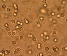
Erythrocytes are disc-shaped structures with a central indentation, the diameter is 4–7 μm . Erythrocytes come in two different forms in urine:
- Isomorphic erythrocytes have the same shape in the urine as the erythrocytes in the blood and usually indicate a urological disease such as kidney tumors , kidney stones or bleeding from the urinary tract (Fig.).
- DysmorphicErythrocytes have irregular shapes and contours and indicate glomerulonephritis (Fig.). Acanthocytes , erythrocytes with vesicle-like protuberances on the cell membrane, show particularly characteristic changes (Fig.). If the proportion of dysmorphic erythrocytes is more than 40% or the proportion of acanthocytes is more than 5% of the erythrocytes counted in the phase contrast microscope, this indicates glomerulonephritis, the patient may then be spared invasive urological diagnostic measures such as a urinary bladder endoscopy (cystoscopy) .
Leukocytes (white blood cells)
The presence of white blood cells in the urine is called leukuria or leukocyturia .
- Neutrophil granulocytes are the leukocytes that are most common in urine. With a diameter of 7–13 μm, they are larger than erythrocytes and can easily be recognized by their granular (granulated) cytoplasm and the lobed cell nucleus (Fig.).
The most common causes for the appearance of neutrophils in the urine are urinary tract infections and admixture of secretions from the genital area to the urine. Other causes include interstitial nephritis , proliferative glomerulonephritis, and urological disorders.
- Eosinophilic granulocytes can be detected in the urine using a special staining method ( Hansel staining , fig.). They occur in a variety of diseases such as acute allergic interstitial nephritis caused by antibiotics as well as rapidly progressive glomerulonephritis , prostatitis , chronic pyelonephritis , schistosomiasis and cholesterol embolism syndrome .
- Lymphocytes appear early in the urine in the cellular rejection of kidney transplants. However, the identification of the cells requires special examination methods which are not available in the routine examination of the urine sediment.
Macrophages (phagocytes)
Macrophages are cells of different sizes, their diameter can be 15 to over 100 μm. The cytoplasm can be filled with fat droplets (Fig.), Vacuoles , granular structures (Fig.) Or entangled (phygocytosed) bacteria. Macrophages occur in the urine in unselective proteinuria , glomerulonephritis, and IgA nephritis .
Renal tubule epithelial cells
Tubular epithelial cells come from the nephron , the canalic system of the kidney. Depending on the tubular segment from which they originate, their diameter varies from 11–15 μm and their shape from rectangular to columnar. It is characterized by a clearly visible cell nucleus with nuclear bodies (nucleolus) (Fig.). Tubular epithelial cells appear in the urine in diseases that damage the nephron, such as acute kidney failure , acute interstitial nephritis , acute renal transplant rejection and, to a lesser extent, proliferative glomerulonephritis .
Urothelial cells
Urothelial cells come from the transitional epithelium (urothelium) , which lines the calyx, renal pelvis, urinary bladder and, in men, the upper urethra. The urothelium consists of several layers.
- Deep urothelial cells: cells from the deep layers are small with a diameter of 13-20 µm, oval to club-shaped. (Fig.).
- Superficial urothelial cells: cells from the superficial layers are larger with a diameter of 20–40 µm. (Fig.).
Deep urothelial cells indicate urological diseases such as bladder cancer , urinary stones or hydronephrosis . In contrast, cells from the superficial layers of the urothelium often occur in urinary tract infections .
Squamous cells
Squamous cells are the largest cells in the urine sediment, their diameter is 45–65 µm (Fig.). They come from the urethra or the external sexual organs (genitals) . In women, the presence of massive amounts of squamous cells in the urine may indicate an infection of the vagina (vaginitis) .

Lipids
Fat droplets (lipids) appear under the light microscope as round, transparent or yellow droplets of different sizes, which can appear either individually, in clumps, in the cytoplasm of macrophages or in cylinders . In the polarizing microscope , fat droplets light up brightly with a dark " Maltese cross " (Fig.). Lipids in the urine can also appear in the form of cholesterol crystals.
An excretion of fats in the urine ( lipiduria ) is typically found in diseases of the kidney corpuscle that are associated with a pronounced protein excretion .
In Fabry disease , lipid droplets can also appear in the urine, but these appear more irregular and show concentric lamellae ( myelin body ) under the electron microscope (Fig.)
cylinder
Cylinders in the urine are cylindrical structures which, as amorphous cylinders, represent an outflow of the renal tubule with Tamm-Horsfall glycoprotein , which is formed in the ascending limb of Henle's loop . A large number of particles can be included in the matrix of Tamm-Horsfall protein, which can indicate various pathological conditions. Due to the mechanism by which these protein cylinders are formed, the trapped particles always come from the kidneys and never from the urinary tract, in the sediment of which they can be detected as hyaline cylinders (in the case of proteinuria ).
The following urine cylinders can be distinguished:
- Hyaline cylinders are colorless and transparent. They can occur in people with kidney disease and normal people (Fig.).
- Hyaline-granulated casts contain granular inclusions in a hyaline matrix and can occur in kidney patients and normal persons (Fig.).
- Granulated cylinders indicate kidney disease.
- Finely granulated cylinders consist of lysosomes containing proteins that have been filtered in the kidney corpuscle (Fig.).
- Coarsely granulated cylinders consist of partially broken down white blood cells (leukocytes) or phagocytes (macrophages) .
- Wax cylinders are wide cylinders with sharp contours and indicate acute or chronic kidney failure (Fig.).
- Fat cylinders usually consist of fat droplets, more rarely of cholesterol crystals, and occur with pronounced protein excretion or nephrotic syndrome (Fig.).
- Erythrocyte casts contain red blood cells (erythrocytes) . They indicate glomerulonephritis , particularly rapidly progressive glomerulonephritis (Fig.).
- Hemoglobin cylinders contain red blood pigment (hemoglobin) . Sometimes a fine granulation is found as remnants of submerged erythrocytes. Hemoglobin cylinders also indicate hemorrhage in the renal corpuscle due to glomerulonephritis , particularly rapidly progressive glomerulonephritis .
- White blood cell casts contain white blood cells (leukocytes) and occur in acute interstitial nephritis and acute pyelonephritis .
- Tubular epithelial cylinders contain epithelial cells from the renal tubules and occur in acute kidney failure , acute interstitial nephritis and proliferative glomerulonephritis .
- Myoglobin cylinders are similar to hemoglobin cylinders. They contain myoglobin and can occur in myoglobinuria due to severe damage to the skeletal muscles ( rhabdomyolysis ).
- Bilirubin cylinders are colored brown and contain bilirubin or its metabolites (metabolites) and occur when red blood cells break down as well as diseases of the liver and biliary tract (Fig.).
- Cylinders that contain microorganisms ( bacteria , yeasts ) can occur with bacterial or fungal infections of the kidneys.
- Crystal cylinders containing uric acid , oxalate or other crystals can occur in all conditions that are associated with increased excretion of crystals ( crystalluria ).
- Mixed cylinders. Finally, mixed forms of different types of cylinders can also occur.
Crystals
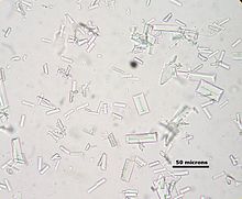
A large number of crystals can appear in the urine , which can often be completely harmless, but can also indicate illnesses or medication taken.

Common crystals:
The excretion of urate, oxalate or phosphate crystals is usually harmless and caused by the precipitation of the substances in concentrated urine. In rare cases, however, crystalluria can indicate metabolic disorders such as hypercalciuria , hyperoxaluria or hyperuricosuria .
- Uric acid crystals are mostly oblong, amber-colored, but can also appear in other shapes. (Fig.). Uric acid crystalluria can lead to acute kidney failure in uric acid nephropathy .
- Amorphous urates consist of irregular granules and can give the urine a red color if they occur in large numbers ( brick dust sediment , fig.).
- Calcium - monohydrate crystals ( Whewellit ) are oval, dumbbell-shaped or bi concave (Fig.). In the context of ethylene glycol poisoning or star fruit poisoning , oxalate crystalluria can lead to acute kidney failure.
- Calcium oxalate dihydrate crystals ( Weddellite ) have the shape of an octahedron (Fig.).
- Calcium phosphate crystals appear as prismatic , star-shaped, needle-shaped and in other shapes (Fig.).
- Triple phosphate crystals ( struvite ) have the shape of a coffin lid (Fig.).
- Amorphous phosphates are found in the form of irregular granules.
Crystals that indicate diseases:
- Cholesterol crystals are thin transparent plates with sharp edges that often clump together (Fig.). Cholesterol crystals occur in the urine with pronounced protein excretion or nephrotic syndrome .
- Cystine crystals appear as irregular hexagonal plates that can be baked together (Fig.). Cystine crystals are evidence (pathognomonic) for the presence of cystinuria .
- 2,8-dihydroxy- adenine crystals are spherical, brownish crystals with stripes extending from the center. The presence of these crystals indicates a defect in the enzyme adenine phosphoribosyl transferase .
Drug Crystals Drug crystals often have atypical shapes.
- A large number of drugs can lead to crystals in the urine, especially in the case of overdose , rapid intravenous administration, lack of albumin , dehydration and depending on the urine pH : sulfadiazine , acyclovir , indinavir , pyridoxylate , primidone , felbamate , amoxicillin and ciprofloxacin .
- Certain drugs can also lead to an increased excretion of calcium oxalate crystals, e.g. B. naftidrofuryloxalate , vitamin C and orlistat .
Guide values in the urine
| Value and unity | |
|---|---|
| Leukocytes | <25 Leu / μl or unit Gpt / l = Giga-Parts per liter |
| Erythrocytes | <2 erythrocytes / μl |
| Squamous epithelia | up to 15 per field of view |
| Round epithelia | no |
| bacteria | no |
| nitrite | 0 mg / dl |
| PH value | 4.6-7.5 |
| protein | <10 mg / dl |
| glucose | 0 mg / dl |
| Ketone | 0 mg / dl |
| Bilirubin | 0 mg / dl |
| Urobilinogen | 0 mg / dl |
| Blood in the urine | negative |
literature
- Giovanni B. Fogazzi et al .: Urinalysis: Core Curriculum 2008 . In: American Journal of Kidney Diseases . Vol. 51, Issue 6, 2008, pp. 1052-1067 ( Article ).
- Giovanni B. Fogazzi, Milan, Italy: Bedside urinary microscopy. Urinary Sediment Part 1: Methods , Part 2: Particles I , Part 3: Particles II , Part 4: Clinical practice I , Part 5: Clinical practice II , Part 6 and last: Contaminants and funny findings . Hands on course from the European Dialysis and Transplantation Organization.
- Lothar Thomas: Laboratory and Diagnosis. Th-Books; 7th edition (November 2007), ISBN 978-3-9805215-6-7
Individual evidence
- ↑ Horst Kremling: On the development of kidney diagnostics. In: Würzburg medical history reports 8, 1990, pp. 27–32; here: p. 27
- ↑ Joseph Loew: About the urine as a diagnostic and prognostic sign. Landshut 1808.
- ↑ G. Guttmann: Technique of the urine examination. Leipzig 1921.
- ↑ Friedrich v. Zglinicki : Uroscopy in the fine arts. An art and medical historical study of the urine examination. Ernst Giebeler, Darmstadt 1982, ISBN 3-921956-24-2 , pp. 1-11 and 17-19.
- ↑ Fig .: Black urine in melanuria
- ↑ Arvin L Santos, et al .: The case: a Caucasian male with dark skin, black urine, and acute kidney injury . In: Kidney International . 76, No. 12, December 2009, ISSN 1523-1755 , pp. 1295-1296. doi : 10.1038 / ki.2009.388 . PMID 19946315 .
- ↑ Horst Kremling : On the development of clinical diagnostics. In: Würzburger medical history reports 23, 2004, pp. 233–261; here: p. 254.
- ↑ CL Foot and JF Fraser: Uroscopic rainbow: modern matula medicine. In: Postgrad Med J. Feb 2006; 82 (964): 126-129. PMC 2596703 (free full text)
- ↑ Geno J. Merli, Howard H. Weitz: The Consult Guys: Green Urine?!?. In: Annals of Internal Medicine. 159, 2013, p. CG3, doi : 10.7326 / G13-3003 .
- ↑ Uma Radha Krishna Pakki Venkata et al .: Quiz Page May 2009: Nephrotic-Range Proteinuria Without Extensive Glomerular Disease . In: American Journal of Kidney Diseases . Vol. 53, Issue 5, 2009, pp. A33-A34 ( article ).
- ↑ M. Lison, SH Blondheim, RN Melmed: A polymorphism of the ability to smell urinary metabolites of asparagus. In: British medical journal. Volume 281, Number 6256, 1980 Dec 20-27, pp. 1676-1678, ISSN 0007-1447 . PMID 7448566 . PMC 1715705 (free full text).
- ↑ K / DOQI: Clinical Practice Guidelines for Chronic Kidney Disease: Evaluation, Classification, and Stratification Part 9. Approach to chronic kidney disease using these guidelines . In: American Journal of Kidney Diseases . Vol. 39, Issue 2, 2002, pp. 215-222 ( online ). Clinical Practice Guidelines for Chronic Kidney Disease: Evaluation, Classification, and Stratification Part 9.Approach to chronic kidney disease using these guidelines ( Memento of March 10, 2015 in the Internet Archive )
- ↑ Fliser, Danilo et al .: Advances in Urinary Proteome Analysis and Biomarker Discovery . In: J Am Soc Nephrol . No. 18 , 2007, p. 1057-1071 ( Article ).
- ↑ Rossing, Kasper et al .: Urinary Proteomics in Diabetes and CKD . In: J Am Soc Nephrol . No. 19 , 2008, p. 1283-1290 ( abstract ).
- ↑ Isomorphic erythrocytes Fogazzi GB, "Urinalysis: Core Curriculum 2008". American Journal of Kidney Diseases 2008; Vol. 51, Issue 6: p. 1052–1067, Supplementary Appendix ( page no longer available , search in web archives ) Info: The link was automatically marked as defective. Please check the link according to the instructions and then remove this notice.
- ↑ Dysmorphic erythrocytes, Fogazzi GB, Urinalysis ( page no longer available , search in web archives ) Info: The link was automatically marked as defective. Please check the link according to the instructions and then remove this notice.
- ↑ Akanthocytes, Fogazzi GB, Urinalysis ( page no longer available , search in web archives ) Info: The link was automatically marked as defective. Please check the link according to the instructions and then remove this notice.
- ↑ Joachim Frey : Diseases of the kidneys, the water and salt balance, the urinary tract and the male sexual organs. In: Ludwig Heilmeyer (ed.): Textbook of internal medicine. Springer-Verlag, Berlin / Göttingen / Heidelberg 1955; 2nd edition ibid. 1961, pp. 893-996, here: pp. 912 f.
- ↑ Neutrophil Granulocytes, Fogazzi GB, Urinalysis ( page no longer available , search in web archives ) Info: The link was automatically marked as defective. Please check the link according to the instructions and then remove this notice.
- ↑ Eosinophilic granulocytes, Hansel staining, from M Kaye, RF Gagnon: Acute allergic interstitial nephritis and eosinophiluria, Kidney International (2008) 73, 980
- ↑ Macrophages with fat droplets, Fogazzi GB, Urinalysis ( page no longer available , search in web archives ) Info: The link was automatically marked as defective. Please check the link according to the instructions and then remove this notice.
- ↑ Granulated macrophage, Fogazzi GB, Urinalysis ( page no longer available , search in web archives ) Info: The link was automatically marked as defective. Please check the link according to the instructions and then remove this notice.
- ↑ Tubular epithelial cell from the proximal tubule, Fogazzi GB, Urinalysis ( page no longer available , search in web archives ) Info: The link was automatically marked as defective. Please check the link according to the instructions and then remove this notice.
- ↑ Deep urothelial cells, Fogazzi GB, urinalysis ( page no longer available , search in web archives ) Info: The link was automatically marked as defective. Please check the link according to the instructions and then remove this notice.
- ↑ Superficial urothelial cells, Fogazzi GB, urinalysis ( page no longer available , search in web archives ) Info: The link was automatically marked as defective. Please check the link according to the instructions and then remove this notice.
- ↑ Squamous epithelial cells, Fogazzi GB, Urinalysis ( page no longer available , search in web archives ) Info: The link was automatically marked as defective. Please check the link according to the instructions and then remove this notice.
- ↑ Macrophage with fat droplets in the polarization microscope, Fogazzi GB, Urinalysis ( page no longer available , search in web archives ) Info: The link was automatically marked as defective. Please check the link according to the instructions and then remove this notice.
- ↑ myelin body Fogazzi GB, urinary sediment
- ↑ Joachim Frey : Diseases of the kidneys, the water and salt balance, the urinary tract and the male sexual organs. In: Ludwig Heilmeyer (ed.): Textbook of internal medicine. Springer-Verlag, Berlin / Göttingen / Heidelberg 1955; 2nd edition ibid. 1961, pp. 893-996, here: pp. 910-912.
- ↑ Hyaline cylinder, Fogazzi GB, Urinalysis ( page no longer available , search in web archives ) Info: The link was automatically marked as defective. Please check the link according to the instructions and then remove this notice.
- ↑ Hyaline-granulated cylinder, Fogazzi GB, Urinalysis ( page no longer available , search in web archives ) Info: The link was automatically marked as defective. Please check the link according to the instructions and then remove this notice.
- ↑ Finely granulated cylinder, Fogazzi GB, Urinalysis ( page no longer available , search in web archives ) Info: The link was automatically marked as defective. Please check the link according to the instructions and then remove this notice.
- ↑ Wax cylinder, Fogazzi GB, Urinalysis ( page no longer available , search in web archives ) Info: The link was automatically marked as defective. Please check the link according to the instructions and then remove this notice.
- ↑ Fat cylinder in the phase contrast microscope, Fogazzi GB, Urinalysis ( page no longer available , search in web archives ) Info: The link was automatically marked as defective. Please check the link according to the instructions and then remove this notice.
- ↑ Fat cylinder in the polarization microscope, Fogazzi GB, Urinalysis ( page no longer available , search in web archives ) Info: The link was automatically marked as defective. Please check the link according to the instructions and then remove this notice.
- ↑ Erythrocyte cylinder, Fogazzi GB, Urinalysis ( page no longer available , search in web archives ) Info: The link was automatically marked as defective. Please check the link according to the instructions and then remove this notice.
- ↑ Bilirubin cylinder, Fogazzi GB, Urinalysis ( page no longer available , search in web archives ) Info: The link was automatically marked as defective. Please check the link according to the instructions and then remove this notice.
- ↑ Uric acid crystal, Fogazzi GB, Urinalysis ( page no longer available , search in web archives ) Info: The link was automatically marked as defective. Please check the link according to the instructions and then remove this notice.
- ↑ Amorphous Urate, Fogazzi GB, Urinalysis ( page no longer available , search in web archives ) Info: The link was automatically marked as defective. Please check the link according to the instructions and then remove this notice.
- ↑ Calcium oxalate monohydrate crystals, Fogazzi GB, Urinalysis ( page no longer available , search in web archives ) Info: The link was automatically marked as defective. Please check the link according to the instructions and then remove this notice.
- ↑ Calcium oxalate dihydrate crystals, Fogazzi GB, Urinalysis ( page no longer available , search in web archives ) Info: The link was automatically marked as defective. Please check the link according to the instructions and then remove this notice.
- ↑ Calcium phosphate crystal, Fogazzi GB, Urinalysis ( page no longer available , search in web archives ) Info: The link was automatically marked as defective. Please check the link according to the instructions and then remove this notice.
- ↑ Triple phosphate crystals, Fogazzi GB, Urinalysis ( page no longer available , search in web archives ) Info: The link was automatically marked as defective. Please check the link according to the instructions and then remove this notice.
- ↑ Cholesterol Crystal, Fogazzi GB, Urinalysis ( page no longer available , search in web archives ) Info: The link was automatically marked as defective. Please check the link according to the instructions and then remove this notice.
- ↑ Zystin-Kristalle, Fogazzi GB, Urinalysis ( page no longer available , search in web archives ) Info: The link was automatically marked as defective. Please check the link according to the instructions and then remove this notice.


