Hematuria
| Classification according to ICD-10 | |
|---|---|
| R31 | Unspecified hematuria |
| N02.- | Recurrent and persistent hematuria |
| ICD-10 online (WHO version 2019) | |
Under a haematuria (from ancient Greek αἷμα haima "blood" and οὖρον ouron "urine") is the frequent occurrence of red blood cells ( erythrocytes ) in urine understood. The two terms erythruria and erythrocyturia are somewhat more precise, but less common . A distinction is made between macrohematuria , in which the blood is already visible to the naked eye as a red coloration of the urine, and microhematuria , which can only be determined by microscopic examination (≥ 3 erythrocytes per field of view under the microscope at 400x magnification). An examination using standard urine test strips cannot distinguish between red blood cells and hemoglobin or myoglobin . Hematuria can indicate diseases of the urogenital tract such as urinary stones , tumors , infections , poisoning , nephritis , congenital malformations of the urinary tract and congenital kidney diseases . Microhematuria with no demonstrable cause is relatively common. Most of the time, especially in young people, it is a temporary and harmless phenomenon. In people over the age of 50, however, a temporary microhematuria can indicate cancer , but in the majority of cases no source of bleeding in the kidneys or urinary tract is found in this age group either.
Macrohematuria
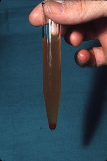
Of gross hematuria is when blood is visible in the urine with the naked eye. The urine appears red or brown in color. Even 1 ml of blood per liter of urine leads to a visible discoloration. In women of childbearing age, mixing with menstrual blood can simulate hematuria. The first step in the investigation of gross hematuria is centrifugation of the urine. This causes the red blood cells to settle in the precipitate. If the supernatant is colorless, there is hematuria. Rarely, with very heavy bleeding or very low urine osmolality , the red blood cells burst and the supernatant turns red. If only the supernatant is red and heme can be detected by a urine test strip , there is either an increased excretion of hemoglobin (hemoglobinuria) or myoglobin (myoglobinuria), the latter in diseases that are associated with muscle breakdown ( rhabdomyolysis ). A red supernatant without evidence of heme can occur with porphyria , after consuming beetroot or taking z. B. rifampicin .
Microhematuria
Microhematuria refers to the detection of blood in the urine by microscopic examination of the urine sediment or by urine test strips if no discoloration of the urine can be seen with the naked eye. In the urine sediment, up to 2 erythrocytes per field of vision are still considered normal at 400-fold magnification.
Other causes of false positive results in the urine test strip are:
- Admixture of seminal fluid ,
- strongly alkaline urine,
- oxidizing substances (e.g. in skin disinfectants)
An excessive intake of vitamin C , on the other hand, can lead to false negative results.
Pathophysiological classification of hematuria
To treat hematuria, it is necessary to locate the source of the bleeding. For this purpose, the division into glomerular and postglomerular hematuria was made.
Glomerular hematuria
In glomerular hematuria, erythrocytes are pressed through damaged basement membranes of the glomerular capillaries and thus damaged. In the phase contrast microscope one can see morphologically changed erythrocytes ( dysmorphic erythrocytes ), which are also known as acanthocytes .
Postglomerular Hematuria
In postglomerular hematuria, the source of the bleeding is behind the glomerulus , so that the erythrocytes that appear in the urine are not pressed through the gaps in the glomerular basement membrane. They are therefore mostly morphologically unchanged ( eumorphic erythrocytes ).
causes
Normally shaped erythrocytes in the urine are usually found in diseases that do not affect the kidney corpuscles, including urinary stones , urinary tract infections and tumors . In patients over 50 years of age, especially in men, a tumor ( kidney cancer , renal pelvic carcinoma , bladder cancer and prostate cancer ) must always be looked for if erythrocytes of normal shape in the urine (normomorphic hematuria) are found. The benign familial hematuria is also known . Sports hematuria can also occur with heavy physical activity .
Other causes of postglomerular hematuria are:
- Kidney cyst with rupture in the hollow system,
- Bladder schistosomiasis ,
- Prostate varices ,
- Blood from the vagina,
- Endometriosis in cyclic hematuria,
- Injuries after inserting a urinary catheter,
- True hermaphroditism with male phenotype as well
- Blood clotting disorders
Dysmorphic erythrocytes, erythrocyte casts and an accompanying evidence of larger amounts of protein in the urine (macroproteinuria) indicate a disease of the kidney parenchyma , usually glomerulonephritis .
Isolated microhematuria, that is, evidence of erythrocytes in the urine without accompanying proteinuria and without erythrocyte casts, is found in 1-4% of the population and around 2.6% of pregnant women. The most frequent cause of an isolated dysmorphic microhematuria is in approx. 2/3 of the cases nonspecific chronic changes of the kidney corpuscles (mostly IgA nephropathy is present), in approx. 1/4 of the cases there is a nephropathy of the thin basement membrane type , and in in the remaining cases no cause can be proven. An increased albumin excretion indicates chronic glomerulonephritis, an increased quotient of immunoglobulin A and C3 is a possible indication of IgA nephropathy.
Because of the lack of therapeutic consequences, invasive diagnostics using kidney puncture are usually not used in isolated dysmorphic microhematuria .
Diagnosis
- Ultrasound examination
- Urinalysis
- Bladder endoscopy ( cystoscopy )
- Gynecological check
- Computed tomography of the kidneys
- iv pyelography
- Retrograde ureteropyelography (if necessary with fluorescence diagnostics and / or flush cytology -> examination for detached tumor cells)
See also
- Hemoglobinuria
- Hematochezia
- Menstruation
- Red urine in newborns: Günther's disease
literature
- S1- Guideline Hematuria - Diagnostic Imaging of the Society for Pediatric Radiology (GPR). In: AWMF online (as of 2013)
Web links
- General Hospital Consilium: Bleeding from the urinary tract
- Hematuria . prostata.de
- Differential diagnosis of hematuria. urologielehrbuch.de
Individual evidence
- ^ Wilhelm Gemoll : Greek-German school and hand dictionary. Munich / Vienna 1965.
- ↑ Gerd Herold and colleagues: Internal Medicine 2020. Self-published, Cologne 2020, ISBN 978-3-9814660-9-6 , p. 601.
- ↑ P. Froom, J. Ribak, J. Benbassat: Significance of microhaematuria in young adults. In: British Medical Journal (Clinical research ed.). Volume 288, Number 6410, January 1984, pp. 20-22, ISSN 0267-0623 . PMID 6418299 . PMC 1444134 (free full text).
- ^ MH Khadra et al .: A prospective analysis of 1,930 patients with hematuria to evaluate current diagnostic practice. In: The Journal of Urology . 163, 2000, pp. 524-527. PMID 10647670 .
- ↑ Willibald Pschyrembel: Clinical Dictionary , 267th edition, de Gruyter, Berlin, Boston 2017, ISBN 978-3-11-049497-6 , p. 710.
- ↑ Horst Kremling : On the development of clinical diagnostics. In: Würzburger medical history reports 23, 2004, pp. 233–261; here: p. 254 f.
- ^ CC Szeto et al .: Prevalence and implications of isolated microscopic hematuria in asymptomatic Chinese pregnant women. In: Nephron. Clinical practice. Volume 105, Number 4, 2007, pp. C147-c152, ISSN 1660-2110 . doi: 10.1159 / 000099004 . PMID 17259739 .
- ↑ P. Shen et al .: Useful indicators for performing renal biopsy in adult patients with isolated microscopic haematuria. In: Int. J. Clin. Pract. 61, 2007, pp. 789-794, PMID 17362478 .

