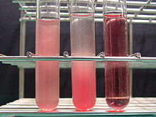
Hemolysis
| Classification according to ICD-10 | |
|---|---|
| D55-D59 | Hemolytic anemia |
| P55-59 | Hemolytic Diseases and Hemolysis in the Newborn |
| ICD-10 online (WHO version 2019) | |

As hemolysis ([ hɛmolyːzə ]; of ancient Greek αἷμα Haima "blood" and λύσις lysis "solution, dissolution, termination"), formerly erythrocyte decay and blood downfall called, refers to the dissolution of red blood cells, the red blood cells . A distinction is made between physiological hemolysis after 120 days and increased hemolysis. The increased hemolysis is associated with a shortened lifespan of the erythrocytes. Anemia occurs if the erythrocyte breakdown exceeds the compensatory new formation.
A naturally increased breakdown takes place immediately after birth , since the fetal erythrocytes then have to be broken down and replaced by erythrocytes for breathing with atmospheric oxygen .
Hemolysis is also a property of microorganisms to break down erythrocytes using hemolysins . This can be proven with the help of blood agar plates . A distinction can then be made on these:
- α-Hemolysis : So-called "greening": The bacteria do not produce any hemolysins, they produce a greenish zone on blood agar, which is not due to real hemolysis, but to discoloration and a loss of potassium in the red blood cells. The reduction in hemoglobin to biliverdin causes the green color. There are still intact erythrocytes. "α-Hemolysis" should not be confused with "α-Hemolysins". The latter are exotoxins (for example from Staphylococcus aureus ) whichcan cause osmotic lysis of a cellby forming a membrane pore . If you are successful with erythrocytes, you actually achieve "β-hemolysis".
- β-hemolysis : extensive dissolution of the erythrocytes and hemoglobin, i.e. real hemolysis. The bacteria produce streptolysin O or S and are surrounded by a clear hemolysis zone, the hemoglobin is completely broken down. In this area all erythrocytes are completely hemolyzed.
- γ-hemolysis : No change in the erythrocytes, so γ-hemolysis is not hemolysis, γ-hemolytic bacteria do not hemolyze.
causes
There is an increased ( pathological ) breakdown of erythrocytes (cell life of less than 100 days)
- in adults:
- mechanical disorders (vascular changes, heart valve prostheses or even extreme marches)
- Membrane defects of the erythrocytes (such as in sickle cell anemia , after exposure to light in erythropoietic protoporphyria )
- from infections such as B. in malaria
- immunological disorders
- Poisons (e.g. from streptococci or certain species of spiders)
- Waldenström's disease
- in newborns: (see phase of life ) significantly increased degradation within a few hours or days
- physiologically through the increased conversion of the fetal to “normal” hemoglobin
- in the case of blood group or Rh factor incompatibilities between mother and child
If more erythrocytes are broken down than are formed, anemia occurs .
When erythrocytes are broken down, bilirubin is produced , so that when the breakdown is increased, bilirubin can also increase, causing jaundice .
Diagnosis
The following laboratory parameters are determined to rule out hemolysis:
- The LDH (the enzyme lactate dehydrogenase ), a marker for cell death. With hemolysis this is usually increased.
- The reticulocyte count ( reticulocytes are young, immature erythrocytes that are increasingly found in the blood when erythrocytes die.)
- Indirect bilirubin, which has not yet been conjugated in the liver, is elevated relatively early in hemolysis.
- Haptoglobin , a protein found in the blood that is used to capture free hemoglobin, may be decreased. It is considered the most sensitive parameter in intravascular hemolytic anemia.
- Free, d. H. Hemoglobin not bound in erythrocytes or haptoglobin in the blood and urine is only found in severe hemolysis.
treatment
In acute cases, transfusions of red cell concentrates may be necessary. Otherwise, the causes should be treated as far as possible.
Normal values of the erythrocyte count and bilirubin in the articles blood test and urine test .
See also
- Myelodysplastic Syndrome
- Hemolysis-in-gel test (HIG test)
literature
- ^ H. Renz-Polster , S. Krautzig, J. Braun: Basic textbook internal medicine. 3. Edition. 2005, ISBN 3-437-41052-0 .
- ↑ Chemgapedia: The alpha hemolysin of the streptococci. Wiley Information Services GmbH.
- ↑ haptoglobin. In: Laborlexikon. Retrieved May 15, 2020 .
