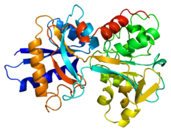
Transferrin
| Serotransferrin | ||
|---|---|---|

|
||
|
Existing structure data: 1a8e , 1a8f , 1b3e , 1bp5 , 1btj , 1d3k , 1d4n , 1dtg , 1fqe , 1fqf , 1jqf , 1n7w , 1n7x , 1n84 , 1oqg , 1oqh , 1ryo , 1suv , 2hau , 2hav , 2o7u , 2o84 |
||
| Properties of human protein | ||
| Mass / length primary structure | 679 AS ; 75.2 kDa | |
| Secondary to quaternary structure | Monomer; 3 glycosyl | |
| Isoforms | several polymorphic variants | |
| Identifier | ||
| Gene names | TF ; PRO1400 | |
| External IDs | ||
| Occurrence | ||
| Homology family | Transferrin | |
| Parent taxon | Euteleostomi | |
| Orthologue | ||
| human | House mouse | |
| Entrez | 7018 | 22041 |
| Ensemble | ENSG00000091513 | ENSMUSG00000032554 |
| UniProt | P02787 | Q921I1 |
| Refseq (mRNA) | NM_001063 | NM_133977 |
| Refseq (protein) | NP_001054 | NP_598738 |
| Gene locus | Chr 3: 133.75 - 133.78 Mb | Chr 9: 103.2 - 103.23 Mb |
| PubMed search | 7018 |
22041
|
Transferrin (from the Latin ferrum 'iron' and transferre 'to transfer ') is a glycoprotein that is produced by the liver and which is mainly responsible for iron transport in vertebrates . It has two binding sites for Fe 3+ ions, binds free iron in the serum and transports it to cells, where it is taken up by transferrin receptors . There are different glycoforms of transferrin, in particular: penta sialo , tetrasialo, trisialo and desialo isoform (CDT). Transferrin is mainly produced in the liver; small amounts are also produced in the testes , brain , spleen and kidneys .

With four percent of the plasma protein, transferrin is the fourth most common protein in the blood plasma. In serum electrophoresis , transferrin runs in the fraction of β-globulins. The iron bound in transferrin amounts to approx. 0.1% of the total iron in the human organism. When fully saturated, the plasma transferrin can absorb approx. 12 mg iron, a comparatively small amount. However, transferrin is still present in the lymph and other body fluids in a similar amount. The transferrin is normally occupied to 30 percent with iron. In the case of iron poisoning, this proportion can rise to 45 percent and therefore the binding capacity of the transferrin can quickly be exhausted, so that free iron is present in the plasma, which is toxic. A very rare metabolic disease, hypotransferrinaemia , is caused by a recessive mutation in the gene coding for transferrin . Arthur L. Schade and Leona Caroline from the Overly Biochemical Research Foundation in New York City are considered the discoverers of transferrin . They published an article about their discovery in Science in 1946 .
Function in iron metabolism
Two transferrin receptors are known to date: Transferrin receptor 1 (TfR1) is expressed in all cells, transferrin receptor 2 (TfR2) mainly in the liver. If iron-laden transferrin binds to one of the receptors, the entire complex (iron, transferrin, receptor) is absorbed via receptor-mediated endocytosis and transported vesicularly to the endosomes . In the early endosomes, the Fe 3+ separates from the transferrin because of the acidic environment .
The transferrin itself (now called apotransferrin) remains bound to its receptor. The receptor / ligand complex is transported to the plasma membrane (recycled) and apotransferrin dissociates from the receptor in the neutral environment of the extracellular fluid. The cycle can begin again. This is a peculiarity insofar as normally in this type of endocytosis either the ligands or even the ligand / receptor complex are broken down by fusion of the vesicle with lysosomes . The transferrin receptors and transferrin reach the cell surface again very quickly via this mechanism. Fe 3+ is transported from the endosome into the cell plasma via a binding protein and is reduced to Fe 2+ in the process. Fe 2+ is then absorbed by ferritins in the cells and oxidized to Fe 3+ .
Iron comes e.g. B. occurs in active centers of enzymes and is important for cell growth. In addition, iron ions are an important part of the oxygen-binding hemoglobin . Iron overload, like iron deficiency, is harmful to the organism. Mutations in TfR2 or in the regulation of TfR1 lead to the clinical picture of hemochromatosis , a harmful iron overload. Iron deficiency can lead to what is known as iron deficiency anemia .
Interpretation of the transferrin level
The normal iron transferrin saturation in adults is around 25–30%. The normal value of the transferrin level (transport iron) in humans is 200–400 mg / dl. An increase in the transferrin level is observed in the presence of an iron deficiency and during pregnancy. A decrease in the transferrin level occurs in the case of chronic inflammation , tumor diseases , iron overload (such as in primary (genetic) or secondary hemochromatosis) or hemolysis . With regular alcohol abuse, the disialo isoform, which usually makes up 1% of total transferrin, increases by up to 10-15 times.
For the detection of liquor , e.g. B. in nasal secretions in a skull base fracture , β2-transferrin can be determined. This isoform is specific for CSF and not contained in the serum. In contrast, β1-transferrin is contained in both CSF and serum.
Soluble transferrin receptor
An increase in this receptor type (sTfR) in the serum correlates very sensitively with a latent iron deficiency.
evolution
The human transferrin belongs to the protein family of transferrins, which not only occur in humans, but homologous genes are also found in other vertebrates and invertebrates. The protein family of transferrins also includes the lactoferrins of mammals.
Transferrine and iron Fe +3 binding protein from Haemophilus influenzae (hFBP) belong to a common protein superfamily. However, the iron-binding structures in transferrins and hFBP emerged from parallel developments .
See also
Individual evidence
- ^ Bowman BH, Yang FM, Adrian GS: Transferrin: evolution and genetic regulation of expression . In: Adv. Genet. . 25, 1988, pp. 1-38. PMID 3057819 .
- ↑ Simon Welch: Transferrin: the iron carrier . CRC Press, Boca Raton 1992, ISBN 0-8493-6793-X .
- ↑ Jacobs EM, Verbeek AL, Kreeftenberg HG, et al. : Changing aspects of HFE-related hereditary haemochromatosis and endeavors to early diagnosis . In: Neth J Med . 65, No. 11, December 2007, pp. 419-24. PMID 18079564 .
- ^ Schade AL, Caroline L: An Iron-binding Component in Human Blood Plasma . In: Science . 104, No. 2702, October 1946, pp. 340-341. doi : 10.1126 / science.104.2702.340 . PMID 17774530 .
- ^ A b Williams, John: The evolution of transferrin. Trends in Biochemical Sciences, Vol. 7, No. 11, 1982, pp. 394-397.
- ↑ Ciuraszkiewicz, Justyna et al .: Reptilian transferrins: evolution of disulphide bridges and conservation of iron-binding center. Gene, Volume 396, No. 1, 2007, pp. 28-38, doi : 10.1016 / j.gene.2007.02.018
- ↑ Liang, Guo Ming, Xun Ping Jiang: Positive selection drives lactoferrin evolution in mammals. Genetica, Vol. 138, No. 7, 2010, pp. 757-762.
- ↑ Bruns, Christopher M. et al .: Structure of Haemophilus influenzae Fe +3 -binding protein reveals convergent evolution within a superfamily. Nature Structural & Molecular Biology, Vol. 4, No. 11, 1997, pp. 919-924, doi : 10.1038 / nsb1197-919
Web links
- Entry on serum transferrin in Flexikon , a wiki of the DocCheck company