
Vaginal vestibule

In human anatomy, the vaginal vestibule ( vestibulum vaginae in Latin ) is the part of the vulva that lies between the labia minora .
In animal anatomy, the area between the vulva and the entrance to the vagina is called the vaginal vestibule. The boundary between the vagina and the vaginal vestibule is defined by the mouth of the urethra ( Meatus urethrae externus ). In addition to the orifice of the urethra, the paraurethral glands (also known as Skene glands) also open ; they release a thin fluid secretion as part of what is known as female ejaculation . The hymen (hymen) is also located on this border .
The spotted hyena has neither a vaginal vestibule nor labia ; in its vagina and urethra - similar to male animals - to form the urogenital canal that runs within the elongated clitoris .
anatomy


Around the vaginal vestibule ( ostium vaginae or vestibulum vaginae ) or vaginal entrance ( introitus vaginae ) are located glands that ensure that the vagina is moistened , as the latter itself has no glands :
- Bartholin's gland ( glandula vestibularis major )
- Paraurethral gland ( Glandula paraurethralis , "Prostata feminina")
- small atrial glands ( glandulae vestibulares minores )
In addition to the smooth muscles of the atrial wall, striated muscles ( musculus constrictor vestibuli ) ensure the closure of the vaginal vestibule. In addition, in some species (humans, dogs, horses) a special swelling tissue ( bulbus vestibuli ) is formed in the lateral atrial wall , which corresponds to the urethral erectile tissue ( corpus spongiosum penis ) of male individuals.
The vestibulum vaginae is the part of the vulva that lies between the labia minora and the entrance to the vagina. The urethral opening, surrounded by the accessory sex glands , such as the outlet ducts of the paraurethral glands (Skene glands), the small atrial glands ( glandulae vestibulares minores ) and the outlet ducts of the two large Bartholin 's glands open into the vestibule . Histologically, it is the zone of transition from the keratinized, multilayered epithelium of the small labia to the non- keratinized epithelium of the vaginal entrance and the vagina ( mucosa ). The urethral opening in turn opens with the ostium urethrae externum directly in front of or above the vaginal entrance, introitus vaginae in the vaginal vestibule ( vestibulum vaginae ) and bulges slightly forward at the anterior wrinkled column of the vagina ( columna rugarum anterior ), this is called bulge or spur-like protrusion Carina urethralis vaginae . While the uncornified squamous epithelium in the vagina is strongly developed, the uncornified squamous epithelium in the vestibulum vaginae is very sensitive (sublime) and has many sensitive nerves and mechanoreceptors . This makes this region easily accessible for sexual stimulation and arousal .
The vestibulum vaginae is delimited towards the inside, i.e. towards the medial side, by the outer part of the hymenal ring; downwards, i.e. towards the anus and laterally, laterally from the so-called Hart line or Hart's line. It is not visible macroscopically . It represents the lateral edge of the vestibulum vaginae and describes ( histologically ) the transition from the non-keratinized squamous epithelium to the slightly keratinized epithelium. It shows the boundary between the “inside” and the “outside world” ( endoderm and ectoderm ).
Diseases
Inflammation of the vaginal vestibule occurs primarily together with a disease of the vagina ( vulvovaginitis ). In terms of differential diagnosis , a rectovestibular fistula ( recto-vaginal auricle fistula , congenitally as atresia ani cum fistula vestibulari with the mouth mostly in front of the posterior commissure of the great labia) ( labia maiora pudendi ) should also be considered.
The Junghundvaginitis is a fairly common condition in domestic dogs before the first heat. It is usually limited to the vaginal vestibule.
literature
- Uwe Gille: Female genital organs. In: F.-V. Salomon et al. a. (Ed.): Anatomy for veterinary medicine. 2nd, expanded edition. Enke, Stuttgart 2008, ISBN 978-3-8304-1075-1 , pp. 379-389.
Web links
Individual evidence
- ↑ Gerald R. Cunha et al: Urogenital System of the Spotted Hyena (Crocuta crocuta Erxleben): A Functional Histological Study. In: Journal of Morpholgy. May 2003, Vol. 256, No. 2, pp. 205-218, PMID 12635111 .
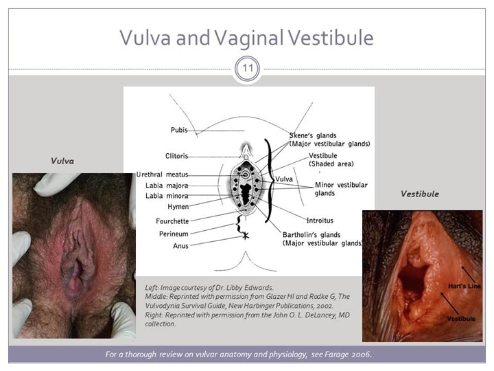
- ^ Vulva and Vaginal Vestibule . The various glands and anatomical structures. From Georgine Lamvu et al: Vulvodynia: An Under-recognized Pain Disorder Affecting 1 in 4 Women and Adolescent Girls - Integrating Current Knowledge Into Clinical Practice. CME / CE, April 2013.
- ↑ Hymen = Virgin Membrane / hymen-virgin-membrane.com formerly known as hymen.pro . Hart's line by D. Berry Hart, AH Barbour: Manual of Gynecology. Edinburgh 1882; "The hymen separates the external genitals from the internal genitals".
- ↑ Manfred Dietel: Pathology: Mamma, Female Genitalia, Pregnancy and Children's Diseases. Springer-Verlag, Heidelberg / New York 2012, ISBN 3-6420-4564-2 , p. 254.
- ↑ Michael K. Hohl , Gudrun Mehring: Painful Vulva: Vulvodynia, Vestibulitis. In: Frauenheilkunde aktuell. 2012, ISSN 1663-6988 , pp. 4–16 ( full text as PDF file ( memento of March 13, 2016 in the Internet Archive )).
- ↑ Günter Thiele (Ed.): Handlexikon der Medizin , Volume 2 (F − K), Urban & Schwarzenberg , Munich / Vienna / Baltimore without year, p. 776.