
Nucleotomy
The discectomy is a surgery for a herniated disc. It is used to remove protruding parts of the intervertebral disc from the spinal cord or spinal nerve canal . A nucleotomy is necessary when nerves are pinched or stressed by the herniated parts of the intervertebral disc and this results in muscle paralysis, sensory disorders of the skin or urinary and fecal incontinence . Whether a nucleotomy is useful for treating pain is disputed among experts.
technology
There are basically two different methods for doing this:
Conventional procedure
In the prone position, in the case of a lumbar disc herniation, a skin incision is made over the spinous processes of the affected movement segment, then the muscles on the affected side are pushed off subperiosteally from the spine . The vertebral arches of the adjacent vertebrae are shown, as well as the ligamentum flavum (“yellow band”) between them. The ligamentum flavum is carefully opened until a punch can be inserted and the space between the vertebral arches can be expanded with it. If necessary, part of one or both vertebral arches must also be removed. When one has penetrated this far, the tensioned root pocket or dural tube appears , which is pulled under with a hook and displaced medially. The herniated disc can now be seen as a plump, whitish, firm protrusion. The tissue is removed with special hollow chisel pliers . As a final check, the free mobility of the root pocket and the dural tube is checked. Hemostasis, irrigation and layered wound closure complete the operation.
Microsurgical procedure
The necessary skin incision is much smaller, a kind of tube (speculum retainer) is inserted, which extends down to the yellow band and the vertebral arches. A surgical microscope is now brought over this tube, the further procedure corresponds to that of the conventional procedure, but with the methods of minimally invasive surgery . The advantage here is the significantly lower trauma, the follow-up treatment time is significantly shorter. With this procedure, the overview that can otherwise be gained during the operation is limited.
Minimally invasive procedure
Here, an optical probe , comparable to an arthroscope , is pushed into the affected root canal under X-ray control and the intervertebral disc tissue is vaporized with a laser guided through this probe .
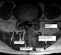
Hemilaminectomy
In this operation, the entire vertebral arch on the affected side was removed. The spinal canal is no longer clearly visible, blood has penetrated here
Nucleotomy
Here the same picture, for a better overview without built-in text. The scar tissue that forms next to the spinous process up to the vertebral arch can be seen very clearly


Endoscopic disc surgery with the Tessys method is also one of the minimally invasive surgical procedures. The surgeon works with special endoscopic instruments and devices through a kind of "natural keyhole" and therefore does not have to destroy the muscle and bone tissue, but just push it to the side. The usual five to ten centimeter long cut on the back is no longer necessary. Instead, the surgeon performs the procedure using a puncture just a few millimeters with a hollow needle. As a result, there are no larger scars, the risk of infection is significantly reduced and the healing process is usually uncomplicated and quick. This enables patients to return to work and everyday life on the same day or after a few days. The success rate of the Tessys method is over 90 percent. In most cases, the surgeon can perform the 45-minute operation on an outpatient basis and under local anesthesia. General anesthesia and the associated risks are therefore not necessary.
Post nucleotomy syndrome
The postnucleotomy syndrome (syn. Postdiskectomy syndrome) is a symptom after intervertebral disc operations. It is characterized by therapy-resistant, sometimes diffuse and burning lower back pain. These are caused by scarring and irritation of the nerve tissue after the operation. Possible causes are cicatricial adhesions between the dural sac, the root pocket and the surrounding bone , postoperative instability with damage to a nerve root, arachnoiditis, somatic and psychosocial effects. If the degeneratively changed intervertebral disc tissue between the vertebrae is removed, the vertebral joints may slip into one another, which is called "telescoping". This leads to chronic, painful wedging of the vertebral joints, which can be avoided in almost any attempt at treatment . Spinal cord stimulation can be used to treat chronic pain .
Individual evidence
- ↑ M. Schubert, A. Helmbrecht, C. Wagner: The microsurgical vs. endoscopic treatment of lumbar disc herniation . In: Orthopedics in Profile . tape 2 , 2010.
- ↑ S3 guideline "Epidural spinal cord stimulation for the therapy of chronic pain". German Society for Neurosurgery eV et al., July 31, 2013, p. Register number 008-023 , accessed on May 15, 2017 .
literature
- M. Schubert, A. Helmbrecht: The transforaminal endoscopic lumbar nucleotomy in all types of herniated discs . In: Surgical General . tape 10 , 2010, p. 519-526 .
- M. Iprenburg, A. Godschalx: Transforaminal Endoscopic Surgery in Lumbar Disc Herniation in an Economic Crisis - The TESSYS Method . In: US Musculoskeletal Review . 2009.
- M. Iprenburg: Transforaminal Endoscopic Surgery - Technique and Provisional Results in Primary Disc Herniation . In: European Musculoskeletal Review . 2007.
- FM Alfen et al .: Developments in the Area of Endoscopic Spine Surgery . In: European Musculoskeletal Review . 2006.