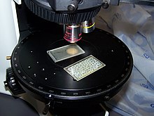
Polarizing microscope
A polarizing microscope is a light microscope that uses polarized light for imaging. It is used to examine optically anisotropic ( birefringent ) objects. These can be crystals or minerals with a corresponding crystal lattice structure (intrinsic birefringence) or isotropic materials on which mechanical forces act (stress birefringence). The third group includes materials that develop birefringent properties due to their arrangement and orientation (shape birefringence in biological or polymeric objects).
In addition to a “normal” light microscope, a polarization microscope contains two polarization filters and a mostly rotating stage. Sometimes so-called compensators are also used to increase the effects (contrasts) or to analyze the strength of the birefringence.
history
In 1808 the French physicist Étienne Louis Malus discovered the refraction and polarization of light. William Nicol invented a prism for polarization in 1829, which was an indispensable part of the polarizing microscope for over 100 years. Later the Nicol prisms were replaced by cheaper polarizing filters.
The first complete polarizing microscope was built by Giovanni Battista Amici in 1830 .
Rudolf Fuess built the first German polarization microscope for petrographic purposes in Berlin in 1875. This was described by Harry Rosenbusch in the yearbook for mineralogy.
Structure and basic principle of the polarizing microscope
Polarizing microscopes usually work in transmitted light mode, although reflected light polarizing microscopes are also available. In transmitted light polarization microscopes, there is a polarization filter , also known as a polarizer or primary filter , below the stage , which linearly polarizes the light from the microscope's light source, i.e. only lets through light that oscillates in one oscillation plane. This direction of oscillation is oriented parallel to the polarizer. Above the stage there is a second polarization filter, which is referred to as an analyzer or secondary filter and is rotated by 90 ° with respect to the first. The direction of oscillation of the previously linearly polarized light is precisely oriented so that it is completely blocked by the analyzer. It does not have any parts that vibrate in the direction of the analyzer. Therefore, the picture appears black. The arrangement of the primary and secondary filters is called " crossed polarizers ".
If there is a sample on the stage between the two polarization filters, the optical conditions can change. Some chemical compounds, for example minerals , have the property of rotating the plane of oscillation of light under certain conditions; they are referred to as birefringent or optically anisotropic . The change in the plane of polarization no longer results in complete extinction - part of the light penetrates the analyzer and the corresponding structures become visible. It is also possible to observe colors occurring due to interference . Optically isotropic materials, however, remain dark.
Rules of extinction
The cancellation rules describe the conditions under which the image is dark:
- Optically isotropic materials never change the direction of oscillation and appear dark regardless of their orientation.
- Optically anisotropic materials are structured in such a way that the light in them can only oscillate in two mutually perpendicular directions. If one of these directions is parallel to the direction of polarization of the exciting light (also called the normal position), the direction of oscillation is retained when the sample is irradiated. Therefore, the light is completely blocked by the analyzer filter. For every anisotropic crystal there are exactly four orientations with extinction due to rotation, all of which are perpendicular to one another.
Lightening and color interference
If an optically anisotropic material is oriented in such a way that the permitted oscillation planes in the crystal are inclined to the plane of polarization of the stimulating light, the light in the crystal is split into two rays with mutually perpendicular planes of polarization (ordinary and extraordinary ray). Certain parts of these are allowed to pass through by the analyzer in the cross position and the image is brightened.
The colored images typical of polarization microscopy are created by interference . In birefringent material the light of the ordinary ray travels at a different speed than the light of the extraordinary ray. When leaving the object, this results in a path difference between the two rays depending on the strength of the birefringence and the thickness of the object. However, as long as the planes of oscillation of the two beams are perpendicular to one another, they cannot interfere with one another. It is only through the analyzer that the components oscillating in the direction of the analyzer are filtered out of both beams. These can intensify or cancel out according to the rules of interference. Since not all wavelengths are equally affected when using white light as excitation , certain color components (wavelength ranges of light) are extinguished and particularly bright and color-intensive images can result. In 1888 , Auguste Michel-Lévy compiled the relationship between thickness, maximum birefringence and path difference of a crystal in a very clear form (Michel-Lévy color scale).

Applications
The polarization microscope is mainly used in mineralogy to examine rock samples. In mineralogy, thin sections are usually created that are radiated through. By examining the various optical properties and colors, conclusions can be drawn about the composition of the rock sample.
Other areas of application are e.g. B. Texture investigations of liquid crystals , investigation of crystal growth, visualization of mechanical stresses (stress birefringence), visualization of crystalline areas (e.g. spherulites ) in polymers, etc. The structure of ice crystals in snow samples can also be investigated with this method. From this, statements about the mechanical properties of the examined snow can be derived, which among other things is relevant for avalanche protection.
Web links
- Description of the polarizing microscope (PDF file; 248 kB)
- Light microscopy online
- Mineral Atlas: Polarizing Microscope (Wiki)
Individual evidence
- ^ Knowledge - Meyers Lexicon: [http://lexikon.meyers.de/wissen/%C3%89tienne+Louis+Malus+(Personen) Étienne Louis Malus] (Meyers Lexicon was switched off).
- ↑ Bergmann-Schaefer: Textbook of Experimental Physics: for use in academic lectures and for self-study. Electromagnetism , Volume 2, Walter de Gruyter, 1999, ISBN 978-3110160970 , page 424 ( online ).
- ↑ History - 19th Century ... (No longer available online.) In: Mikoskop-Museum.de. Olympus Deutschland GmbH, archived from the original on February 15, 2009 ; Retrieved January 6, 2009 . Info: The archive link was inserted automatically and has not yet been checked. Please check the original and archive link according to the instructions and then remove this notice.
- ↑ Leopold Dippel, The microscope and its application .
- ^ Museum of optical instruments: Polarizing microscope according to Rosenbusch .
- ↑ Brochure from Zeiss Amplival pol polarization microscope (PDF; 2.0 MB), accessed on February 26, 2014.


