
Thin section

A thin section is a solid preparation for microscopic examination. The examined materials are predominantly rocks, ceramics and all materials which are transparent in thin layers, but which cannot or only poorly be cut accordingly thin, for example wood, bones and teeth.
Rocks , soils and ceramics are almost invariably opaque and therefore cannot easily be examined under a transmitted light microscope . Only from a thickness of 0.03 to 0.02 mm (30 to 20 µm) does a sample of these materials allow sufficient light to pass through so that they are suitable for transmitted light microscopy. The examination is carried out in normal and polarized light as well as with special wavelengths (e.g. UV light).
history


The method was developed in the first half of the 19th century by several scientists independently of one another, whereby the earliest attempts can be traced back to the English physicist William Nicol , who made such preparations as early as 1831 and also published a description of the process. But it was only the English naturalist Henry Clifton Sorby who systematically applied the method to rocks and published an article in the Journal of the Geological Society in London in 1858 .
In the field of palaeontological research, the geoscientists Franz Unger and Carl Ferdinand Peters made early achievements with thin-section microscopy . In 1842, Unger described his method of using thin sections when examining fossil woods. Around 1840 he had made finite thin sections of such objects on behalf of Stephan Ladislaus . Peters carried out (published) thin sections around 1855 in order to obtain further information for taxonomic assessments.
Ferdinand Zirkel made a decisive contribution to the dissemination of this research method in the geosciences , who presented it on February 3, 1863 at a meeting of the Imperial Geological Institute and who had previously learned from Sorby. After that, the method quickly found its way into the practice of petrography . The first textbooks on the subject were published in 1873 by Ferdinand Zirkel and Karl Heinrich Rosenbusch .
In the 19th century, the representation of thin-section images was a problem that could only be solved in an unsatisfactory manner for the specialist literature, because the printing techniques of the time did not enable an authentic reproduction of the often demanding structural images. For this reason, it was at times common to create corresponding thin-section images by drawing with subsequent hand coloring and to implement them in printing with the means of lithography .
Manufacturing
Geological and technical preparations

To produce a thin section of stone or ceramic, a 0.5-1.0 cm thick disc is sawn off the sample using a diamond-tipped saw blade. The pane is sanded by hand on one of the cut surfaces and fixed with this surface on a thin glass plate using a relatively easily soluble adhesive. The glass plate with the sample is then clamped in a precision grinding machine and the sample is ground from the other side until it is completely flat. After the glass plate with the sample has been removed from the grinding table, the flat surface is glued to a glass slide using a special synthetic resin (in the early years with Canada balsam ) . The now almost finished preparation is removed from the glass pane after the resin has hardened. The slide is clamped in the precision grinding machine and the sample is now ground down to the desired thickness (standard thickness for mineralogical thin sections is 25 µm) from the other side, which was previously attached to the glass pane .
Technical granular preparations or unstable sedimentary rocks (e.g. many types of quartz sandstone ) are first poured into synthetic resin or impregnated with synthetic resin and, after the resin has hardened, further processed like the above-mentioned solids.
Soil preparations
To produce a thin section of the soil , an undisturbed soil sample using Kubiëna boxes (named after WL Kubiëna, who with his micromorphological studies on soils carried out since the 1930s, undoubtedly pioneered the field in this area) and modifications of these boxes is used. If extraction with Kubiëna boxes is not possible due to a high stone content or larger artifacts, larger frames can be used or the sample can be coated with plaster. The soil samples are finally dried in the laboratory (air drying, freeze drying or drying over acetone ). They are then poured into synthetic resin in a vacuum cabinet (so that no air bubbles remain in the samples), similar to granular technical samples or sediment samples . After the resin has hardened, proceed as described above .
For some more specific examinations (micro sample etc.) uncovered samples are necessary. The sample can also be covered with a cover slip. For examinations z. For example, polished, uncovered sections are required on the microprobe; this requires special methods that do not create a relief.
- Polarizing microscopy: parallel vs. crossed polarizers

Thin section of a garnet - mica slate under the microscope at linearly polarized light
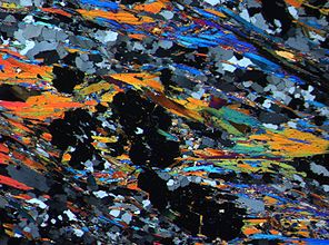
The same image section as in the adjacent image with crossed polarizers


Medical preparations
In bone pathology , thin sections are used for the microscopic examination of biological hard tissue ( bones and teeth ) and especially for assessing the healing of metal endoprostheses (for example hip joint prostheses or dental implants ). Experimental studies with systematic microscopic examinations of rows of sections at the interfaces between bone tissue and implant can significantly improve the surfaces of endoprostheses so that the implant is optimally accepted by the body.
For the production of bone tissue thin sections , the bone tissue is fixed and then embedded in PMMA resin (Plexiglas) without decalcification . After hardening, the entire plastic and tissue block can be sawn thin and then sanded down to a thickness of µm until it is transparent and colored if necessary. With the polarization microscopy of these tissue thin sections, especially the collagen fiber bundles oriented along the force vectors can be made clearly visible without histological staining .
Rock examinations using thin sections
When examining rock samples , a distinction is made between qualitative and quantitative goals. Furthermore, general characteristics are the purpose of consideration.
General goals
- spatial orientation of crystals and grain aggregates
- Distribution character of minerals in rock
- Grain or crystal forms
- Grain bonds
Qualitative targets
- Determination of the rock-forming minerals
- Composition of mixed crystals using optical data
- Degradation processes and their achieved status in crystalline structures (weathering)
Quantitative goals
- Ratios of various minerals in the rock
- Grain size distribution
Results and interpretation in soil science
Before you can interpret a thin section, it is first necessary to describe this thin section and further thin sections of other samples that are directly or indirectly related to it (if any) in detail.
In soil science and archeology , among other things, the following are recorded:
- the microstructure (aggregates, cavities, passages),
- the so-called basic mass (i.e. the organic and mineral fine and ultra-fine material),
- the organic material not incorporated into the base mass and
- the individual soil characteristics and peculiarities.
To characterize these components, among other things, size, shape, texture, variability, frequency, color, translucence, relationship and position of the components to each other and any resulting patterns are described.
The more or less pronounced characteristics and combinations of characteristics visible in the thin section of the soil are a “snapshot”: They reflect the development of a soil and the processes in it up to the point of sampling. From an archaeological point of view, charcoal residues, bone fragments, particles of burnt clay, slag and ore residues, excrement, eggshells, herringbones, etc. are of particular interest, because - depending on the position in the profile - ideally one can use the "microscopic finds" in thin section ( in connection with other thin sections of the same finding as well as possibly existing "macroscopic finds") reconstruct the history of a finding: From the former use or function of an object itself (for example of mine houses), through the environmental conditions (e.g. animal husbandry) to for backfilling an object as well as the origin and composition of this backfill material, which can document human activities in the vicinity of the finding.
Related methods
If opaque materials are only to be examined in incident light, one-sided ground and polished surfaces on the object, so-called bevels, are sufficient.
Even within metallography , one occasionally works with micrographs .
The methods used in medical research to produce soft tissue sections (hard tissue containing calcium carbonate must first be decalcified) for transmitted light microscopy are described under Histology and Microtome .
literature
- Arnd Peschel: Natural stones. German Publisher f. the basic industry, Leipzig 1977.
- Hans Pichler, Cornelia Schmitt-Riegraf: Rock -forming minerals in thin sections. Ferd. Enke Verlag, Stuttgart, 1993, ISBN 3-432-95522-7 .
- DL Rowell: Soil Science. Investigation methods and their applications. Springer, Berlin / New York, 1997, ISBN 978-3-540-61825-6 .
Web links
- The production of a thin section. on www.geo.tu-freiberg.de
- Manufacture of rock thin sections. at www.titan.minpet.unibas.ch
Individual evidence
- ↑ a b H. v. Philipsborn: The historical development of microscopic methods in mineralogy and their significance for general microscopy and for technology . In: H. Freund (Hrsg.): Handbuch der Mikoskopie in der Technik . tape IV / 1 . Umschau, Frankfurt 1955, p. 12-17 .
- ↑ Emanuel Bořický: The work of the geological department of the regional exploration of Bohemia, Part II, Petrographic studies on the basalt rocks of Bohemia . Prague (Řivnač) 1873, pp. 3–4.
- ^ Bernhard Hubmann: Paleontological thin section investigations in Austria-Hungary before 1860 by CF Peters and F. Unger . In: Abhandlungen der Geologische Bundesanstalt Vol. 56/1 (1999), pp. 171–176. ISSN 0378-0864 .
- ↑ DW Humphries: Methods of thin section production . Enke, Stuttgart 1994, ISBN 3-432-26091-1 , p. 20 .
- ↑ a b R. K. Schenk, W. Herrmann: Histological investigations on the healing of cementless implants. P. 51–57 in: E. Morscher: The cementless fixation of hip endoprostheses. Springer, 1983, ISBN 978-3-662-00968-0
- ↑ K. Donath: The Diagnostic Value of the New Method for the Study of Undecalcified Bones and Teeth with Attached Soft Tissue, (Säge-Schliff, (Sawing and Grinding) Technique). Pathology - Research and Practice. Vol. 179, 1985, pp. 631-633, doi: 10.1016 / S0344-0338 (85) 80209-0
- ↑ NM Meenen, W. Flosdorff, M. Dallek, K. Donath, KH Jungbluth: hydroxyapatite great for subchondral bone replacement joints - An animal optical polarization study. Pp. 271–275 in: Hans-Jürgen Pesch, Hartmut Stöß, Benno Kummer (Eds.): Osteologie aktuell VII (7th annual meeting of the German Society for Osteology eV, March 26-28, 1992 in Erlangen). Springer-Verlag, ISBN 978-3-540-56630-4