
Quadriceps tendon rupture
| Classification according to ICD-10 | |
|---|---|
| S76.1 | Injury to the muscle and tendon of the quadriceps femoris muscle |
| ICD-10 online (WHO version 2019) | |
The quadriceps tendon rupture is a tear in the tendon of the large extensor muscle ( rectus femoris muscle ), usually directly at its attachment to the kneecap ( patella ) or a few centimeters above.
causes


The rupture of the quadriceps tendon is usually due to an over-tension trauma against resistance or a strong tension in the flexion of the knee joint . Elderly patients with degenerative (hyaline degeneration) changes in the tendon are mainly affected . Occasionally, metabolic and circulatory disorders , repeated "microtraumas" and - occasionally - repeated injections into the tendon are also responsible.
Diagnosis
The typically localized pain, the inability to stretch the knee joint against resistance, and the usually palpable gap in the tendon make the injury usually clinically clear. In cases of doubt, for example if the examinability of the knee joint is limited by severe swelling and pain, sonography or magnetic resonance imaging ( MRI or magnetic resonance imaging ) can provide clarity. A palpable gap in the tendon with retained extensibility indicates an incomplete rupture that often does not require surgical treatment.
therapy
The treatment for the complete rupture is usually the tendon suture. Here, the seams are usually guided through drill channels made in the kneecap, since the crack is often directly at the kneecap attachment or just above it. The follow-up treatment consists of the primary immobilization of the knee joint in a cast or now more often in an orthosis . With the latter, the flexion of the knee joint can be released step by step to a defined extent after four to six weeks. The release of flexion after six to ten weeks is followed by an intensive physiotherapy exercise treatment, which on the one hand strengthens the thigh muscles, which are now atrophied , and on the other hand promotes mobility in the knee joint.
forecast
With consistent primary and follow-up treatment, the prognosis is favorable; in most cases, full restoration of the function of the extensor apparatus can be achieved. The result can be jeopardized by releasing flexion too early, but also by postoperative wound infections. Depending on the initial situation, however, renewed ruptures of the tendon may occur, particularly in patients with previous degenerative damage.
More picture examples

Complete tear with a deep patella

Partial tear of the tendon, which is also visible on the x-ray
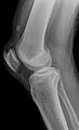
Tear of the tendon with previous damage with tendon attachment calcifications that are now pulled up


See also
swell
- A. Rüter, O. Trentz, M. Wagner (Ed.): Trauma surgery. Study ed. the 2nd, revised. u. exp. Edition. Urban & Fischer, Munich 2008, ISBN 978-3-437-21851-4 , p. 1103.