Achilles tendon rupture
| Classification according to ICD-10 | |
|---|---|
| S86.0 | Achilles tendon injury |
| ICD-10 online (WHO version 2019) | |
The Achilles tendon rupture (ASR) describes the covered (without skin damage) tear of the Achilles tendon . It occurs when the calf muscles are suddenly tensed during exercise or other physical activities. Most patients are in their 4th and 5th decades of life. The rupture can be treated surgically or conservatively with a solid bandage.
Anatomical-physiological basics

The Achilles tendon - Tendo calcaneus (Achilles) - is the strongest tendon in the human body. She sits on the heel bone hump ( calcaneal tuber ) and incorporated as a common tendon of the calf muscle ( triceps surae ) the terminal tendons of the three calf muscles (soleus and Mm. Gastrocnemius medial and lateral).
It can withstand loads of 60 to 100 N / mm², which corresponds to a load capacity of up to 800 kilograms for an area of 80 mm². A rupture of the Achilles tendon when the triceps surae muscle is suddenly tensed usually only occurs when there has been previous damage due to excessive or incorrect loading.
The tendon repeatedly experiences minor injuries that disrupt the blood supply to the tissue and thus lead to the degeneration of the tissue. These changes are most pronounced in an area 2 to 6 cm above the insertion (so-called "Achilles tendon waist"), where the tendon is poorly supplied and where the tear usually also occurs. The tendon then suddenly breaks with a loud, whip-cracking sound. The plantar flexion or toe walking is then only a very limited extent.
The mechanical stability of the Achilles tendon can be trained, as has been proven experimentally. The improved stability, however, regresses when the physical activity is reduced. Therefore, Achilles tendons in young adults with a high volume of training are particularly stable against tensile stress. If there is a sport break in the course of further life due to work or starting a family, the stability decreases again. If the old, usual sporting activities are suddenly resumed in middle age (40+), the risk of suffering an Achilles tendon rupture is particularly high. The position of the foot in relation to the lower leg also plays a role: With supination and pronation (lateral tilting of the rear foot in the lower ankle), the flat Achilles tendon is subject to edge loads that quickly exceed the maximum load capacity of the entire straight tendon and thus at the point the greatest force lead to the primary tear, which is then propagated (forwarded) over the entire width of the tendon. This is why typical Achilles tendon tears do not occur when walking straight, but especially when the feet are in an inclined position, such as in tennis or soccer.
etiology
A rupture of the Achilles tendon is usually a sudden severing of the Achilles tendon. The Achilles tendon rupture rarely occurs with prior notice, for example due to Achilles tendon pain or irritation. As a result, people who are physically active are more often affected by an Achilles tendon rupture. Sports that are characterized by rapid changes of direction, such as soccer or handball , and which place such high stress on the Achilles tendon, are at the highest risk of an Achilles tendon rupture. About 89 percent of all Achilles tendon ruptures can be attributed to these indirect trauma . There is an increase in active men between the ages of 30 and 50.
Another rare cause of a spontaneous Achilles tendon rupture is the use of an antibiotic from the group of gyrase inhibitors , such as levofloxacin . The elderly and those taking corticosteroids or other immunosuppressants are at greater risk. This is explained by an increased expression of matrix metalloproteinases , which reduce the strength of the tendons.
Open transections of the Achilles tendon are also rare, as they occur when the heel region comes into contact with sharp-edged metal sheets, glass panes or z. B. garden instruments can occur. Due to the wound, there is a higher risk of infection and, as accompanying injuries, also vascular or nerve lesions or further tendon cuts. This injury must only be treated surgically and urgently. Further treatment requires a somewhat longer immobilization and later resumption of full function, because the sharp incision has a worse healing tendency than the covered spontaneous rupture.
Incidence
The tear of the Achilles tendon is a rather rare sports injury. In a study carried out from 1972 to 1997, the proportion of Achilles tendon injuries was around 2 percent (701 of 34,742 recorded sports injuries). The number of cases increased significantly in Germany towards the end of the 20th century. This is mainly due to new types of sport and increasing obesity in the population. In 1996 there were 15,000 to 20,000 cases of Achilles tendon rupture in Germany. For men, the incidence in Scotland is 6.3 to 7.3 per 100,000 population. For women, the value is 3 to 4.7 per 100,000 inhabitants. The incidence increased significantly between 1980 and 1995. The frequent resumption of highly active sport in the age 35+ typical for Achilles tendon ruptures after a work-related or family-related break plays a role here.
Diagnosis
Clinically, with very fresh ruptures, there is usually a palpable gap, most often a few centimeters above the insertion on the heel bone (calcaneus) . After a while, the area will be bloodshot and swollen. Walking on tiptoe is no longer possible. In the supine position, the foot can no longer be moved plantar (towards the sole of the foot). A residual function can be simulated by the tendon of the M. tibialis posterior and by the long toe flexors. If the rupture is complete, the Thompson test is positive: the patient lies on his stomach, his feet hang over the edge of the examination table. If the calf muscle is now squeezed from both sides, the foot is normally plantar flexed, but not in the case of an Achilles tendon rupture. A residual function can be retained through the function of the plantaris longus, but this should not be misinterpreted.
The rupture can be detected by ultrasound. The advantage of sonography is the possibility of dynamic examinations under functional stress. On the basis of the examination results, a decision can be made on the therapy method with the distance between the two tendon ends: Only a short distance increases the probability of success of a functional-conservative treatment. Surgical procedures are preferred when the distance is great.
A magnetic resonance examination is carried out today for all findings that cannot be reliably assessed even with ultrasound. Fresh, but also outdated ruptures, degenerations such as calcium deposits and partial ruptures can be detected here. Magnetic resonance imaging provides essential information, which often also has operative consequences, particularly in the case of atypical anamnesis (without adequate trauma) and also in the context of treatments with unclear processes.
treatment
In principle, a conservative or surgical approach can be used in the event of a crack. The decision depends on the age, the risk factors (smoking, arterial circulatory disorders, obesity , medication intake) and the athletic demands of the patient. While surgical treatment was previously given priority in physically active patients, this view has been increasingly weakened by recent studies in the last two decades. So shows z. B. an English study from 2014 that the risk of a new crack can be reduced to 1.1% even with conservative treatment through an appropriate management program. In the study, competitive athletes were also treated conservatively. In previous studies without adequate management, RE rupture rates of 8 to 20% for conservative therapy and 3 to 6% for surgery were given. The conservative treatment is also equivalent to the operation for the other results such as mobility and strength in the lower leg, which are measured in the so-called AS score. With regard to possible other complications (problems with the surgical scar, infections, nerve injuries), conservative treatment is judged to be clearly advantageous.
Treatment without surgery (functionally conservative)
The foot is immobilized by a firm bandage (e.g. plaster of paris, special shoe, orthosis ) for about 2 weeks in the equinus position. The healing progress can e.g. B. be monitored with ultrasound examinations. This is followed by the provision of a special boot in which the heel elevation can be adjusted in stages from 30 degrees up to for about six weeks. According to more recent studies, the rate of re-ruptures in conservative therapy can be reduced to the level of surgical treatments. The prerequisite for this is early exposure. Due to the minor complications, conservative therapy is recommended if the distance between the two torn ends is less than 10 mm.
Operative treatment
Both the open operation and the minimally invasive procedure are performed either under local anesthesia , regional anesthesia or general anesthesia (general anesthesia ). A pressure cuff on the thigh can cut off the blood supply for the duration of the procedure (tourniquet) in order to reduce blood loss and improve the overview of the operating area. With all risk factors related to blood circulation (diabetes, smoker, age, arterial circulatory disorders) the tourniquet should not be used, as there is a high risk of wound healing disorders in the ankle area. Much of the criticism of surgical methods can be traced back to these circulation-related healing disorders. The rate of complications can be significantly reduced by dispensing with tourniquets and by carefully preparing the tissue and using simple suturing techniques.
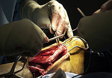
- Open operation: The torn and separated tendon ends are exposed and reunited by a suture and / or adhesive. Simple suture techniques such as lace sutures / braiding according to Bunnell or the modified Kessler-Kirchmayr tendon sutures are used. If the tendon is out of date or the tendon is severely frayed, tendon plastic surgery must be performed to bridge Achilles tendon defects. T. Neighboring tendons are used to strengthen the Achilles tendon (tendon plastic through an interposal ). The functionally insignificant Plantaris longus tendon, which runs parallel to the Achilles tendon and is applied in more than 70% of all patients, is particularly suitable. With a special tendon stripper, an approximately 15 cm long part of the tendon is removed up to the calf, leaving the tendon on the heel bone. The tendon is braided with a special needle following the usual suture. This form of restoration achieves the highest stability values for the seam.
In the case of a rupture of a degeneratively changed tendon very close to the bone, it must be reattached directly to the heel bone. Bone anchor sutures are used for this today.
In the rare bony tearing of the tendon directly from the heel bone, the piece of bone is screwed back onto the calcaneus cusp together with the tendon attached.
For outdated ruptures with defect healing, so-called overturning plastics are performed. A strip with a distal stalk is cut proximally from the muscle fascia of the triceps surae muscle (calf muscle), which serves the Achilles tendon. This fascia strip is folded (overturned) over the defect zone in the direction of the heel bone and fixed there with absorbable sutures. Follow-up treatment must be carried out more carefully than with normal tendon sutures. In any case, risk behavior should be avoided (smoking).
- Minimally invasive surgery : In contrast to open surgery, the operating area is not exposed, but the torn tendon ends are found through small incisions and connected to each other again. A small cross-section at the level of the tear site makes it possible to control the approach of the torn tendon ends. A major complication here is the lesion of the sural nerve with a corresponding loss of skin sensitivity in the foot area when the covered suture encompasses the nerve. The advantage of the minimally invasive method is the low risk of wound healing disorders.
Depending on the findings of the operation, it may be necessary to insert a wound drain to suck off wound secretion and blood.
Even after an operative treatment, it is necessary to immobilize or protect the leg for several weeks. To relieve the tendons, the foot is initially fixed in the equinus foot position (around 30 ° to 40 °) and then slowly returned to its normal position over a period of weeks. Close controls by the doctor treating you are necessary.
An adapted dose of heparin is necessary for the prophylaxis of a thrombosis that is common in foot and lower leg injuries. If the patient is fully loaded, even within a immobilizing orthosis , if risk factors (smoking, contraceptive hormone treatment) are excluded, drug-based thrombosis prophylaxis can be dispensed with.
Spontaneous course (non-treatment)
If an Achilles tendon rupture is not recognized or not treated, a regenerated tendon (neo-tendon) forms within two to four months, which only enables a powerless function in the ankle. The loss of strength is caused by the considerable lengthening of the tendon due to the regenerated scar, so that the calf muscle cannot work in the optimal range of strength if the tendon is extended by several centimeters. In this case, despite intensive training, the old tension relationships between muscles and tendons cannot be restored. Similar courses are observed when conservative therapy fails and operations have failed. This can be seen in a significant atrophy of the calf muscles , loss of strength and a limp or dragging of the affected foot.
rehabilitation
With both approaches, the tendon is usually immobilized in a lower leg cast or in an orthosis for six weeks . After that, a gradually graduated rehabilitation program begins .
During this time, the foot is held in the equinus position (about 10 ° to 20 °), which can be slowly reduced depending on the therapy. The risk of another crack is particularly high in the 8th to 10th week. Loose running training on a level surface can usually be started after about four to six months.
The healed Achilles tendon - like all other torn tendons - never regains its full load capacity. However, the increase in diameter of the healed tendon, which is always observed, results in sufficient stability per cross-sectional unit. With an optimal course it is up to 90% of the healthy tendon. In the optimal case, sport can be practiced again without restrictions - up to competitive sport. The risk of a new rupture of the Achilles tendon ( relapse ) is 1 to 2 percent when a therapeutic measure is used. and thus about the same as the risk of injury from a healthy Achilles tendon.
Web links
- Guideline Achilles tendon rupture of the German Society for Orthopedics and Orthopedic Surgery. (PDF; 37 kB) DGOOC online (not updated)
Individual evidence
- ↑ a b c d J. Isbach: The operative treatment of the Achilles tendon rupture taking into account the treatment results from the years 1992 to 1997. (PDF; 1.9 MB) Dissertation . University of Münster, 2007.
- ^ H. Vyas, G. Krishnaswamy: Quinolone-associated rupture of the Achilles' tendon. In: New England Journal of Medicine. 357, 2007, p. 2067. PMID 18003963 .
- ↑ K. Steinbrück: Achilles tendon rupture in sport - epidemiology, current diagnostics, therapy and rehabilitation. In: German magazine for sports medicine. 51, 2000, pp. 154-160.
- ↑ H. Lill: Current status of the treatment of Achilles tendon ruptures surgeon. 67, 1996, pp. 1160-1165.
- ↑ M. Majewski et al. a .: The fresh Achilles tendon rupture: A prospective investigation to assess various therapy options. In: Orthopedics. 9, 2000, pp. 670-676. PMID 10986713 .
- ↑ N. Maffulli et al. a .: Changing incidence of Achilles tendon rupture in Scotland: a 15-year study. In: Clin J Sports Med. 9, 1999, pp. 157-160. PMID 10512344 .
- ↑ M. Gebauer, FT Beil, J. Beckmann, AM Sárváry, P. Ueblacker, AH Ruecker, J. Holste, NM Meenen: Mechanical evaluation of different techniques for Achilles tendon repair. In: Arch Orthop Trauma Surg. 2007 Nov; 127 (9), pp. 795-799.
- ↑ AM Hutchison, C. Topliss, D. Beard, RM Evans, P. Williams: The treatment of a rupture of the Achilles tendon using a dedicated management program . In: Bone Joint J . 97-B, no. 4 , April 1, 2015, ISSN 2049-4394 , p. 510-515 , doi : 10.1302 / 0301-620X.97B4.35314 , PMID 25820890 ( org.uk [accessed March 25, 2017]).
- ↑ Hao Zhang, Hao Tang, Qianyun He, Qiang Wei, Dake Tong: Surgical Versus Conservative Intervention for Acute Achilles Tendon Rupture . In: Medicine . tape 94 , no. 45 , November 13, 2015, ISSN 0025-7974 , doi : 10.1097 / MD.0000000000001951 , PMID 26559266 , PMC 4912260 (free full text).
- ↑ a b H. Thermann u. a .: Achilles tendon rupture. In: Orthopedist. 29, 2000, pp. 235-250. PMID 10798233 (review).


