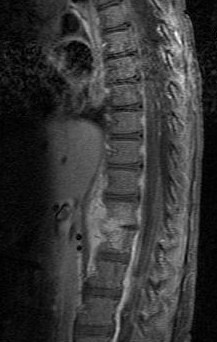
Spondylodiscitis
| Classification according to ICD-10 | |
|---|---|
| M46.49 | Discitis, unspecified |
| ICD-10 online (WHO version 2019) | |

The Spondylodiscitis or osteomyelitis of the vertebrae is inflammation of the intervertebral disc and the two adjacent vertebral bodies , which, as well as inflammatory rheumatic diseases is mostly caused by bacterial infections.
Symptoms
The symptoms are initially unspecific, the pain corresponds in intensity and extent to those that can also be caused by degenerative changes in the spine. Pain occurs mostly at night or during exercise and is sometimes accompanied by night sweats, fever, or weight loss. Strong knocking and pressure pain in the affected vertebrae is very characteristic.
Pathogen
In spondylodiscitis, a distinction is made between community-acquired infections and nosocomial infections, which may occur after neurosurgical interventions or catheter-associated with central venous catheters or dialysis catheters, each with a different spectrum of pathogens.
By far the most common cause of an intervertebral disc infection is Staphylococcus aureus , rare are Staphylococcus epidermidis , viridans streptococci , Escherichia coli , Pseudomonas aeruginosa , Pneumococcus , Clostridium perfringens , Proteus mirabilis , Haemophilus aphrophilus , Mycobacterium tuberculosis , leprae Mycobacterium and Veillonella parvula .
diagnosis
The laboratory data can provide an indication of an inflammatory process, but can also remain inconspicuous. In the X-ray image, the intervertebral disc tissue appears to be compressed in the later stage, and at the beginning there are often no pathological findings. Magnetic resonance imaging often provides information , and computed tomography can also provide information. Evidence of the pathogen may be possible through blood cultures . In addition, a puncture can be performed for a more precise determination of the pathogen .
therapy
Depending on the severity of the clinical picture, the inevitable, intensive antibiotic treatment may have to be supplemented by surgical measures. An operation is especially necessary if a calculated antibiotic does not lead to improvement or neurological deficits occur. The intervertebral disc tissue is surgically removed and the adjacent vertebral bodies are splinted together to prevent any movement in the affected segment. A CT-guided biopsy can sometimes be used to confirm the pathogen and to determine resistance.
Those affected have to stay in bed for 2 to 8 weeks, even with conservative treatment. This is also strongly dependent on the severity of the inflammation, but also on the age of the person affected. In younger people, the therapy is often faster and they almost regain their mobility. A stabilizing trunk orthotic is also given. The necessary treatment and monitoring of the course usually take a long time, usually well over a year.
The inflammation can cause the entire or part of the intervertebral disc to dissolve. The two vertebral bodies then grow together and ultimately the same thing happens as in a surgical procedure.
literature
- Sobottke, R. et al .: Current diagnosis and therapy of spondylodiscitis . In: Dtsch Arztebl . No. 105 (10) , 2008, pp. 181-187 ( Article ).
- S. Rajasekaran: History of spine surgery for tuberculous spondylodiscitis . The trauma surgeon 118 (2015), pp. 19-27.
- Marianne Abele-Horn: Antimicrobial Therapy. Decision support for the treatment and prophylaxis of infectious diseases. With the collaboration of Werner Heinz, Hartwig Klinker, Johann Schurz and August Stich, 2nd, revised and expanded edition. Peter Wiehl, Marburg 2009, ISBN 978-3-927219-14-4 , pp. 167-170 ( spondylodiscitis or osteomyelitia of the vertebral bodies ).
Individual evidence
- ↑ Marianne Abele-Horn (2009), p. 167 f.