Internal total reflection fluorescence microscopy
Total internal reflection fluorescence microscope ( English reflection fluorescence microscopy total internal , TIRFM) is a particular method of light microscopy , in which the fluorescence of a specimen on a evanescent is excited (decaying) field. To generate the evanescent field, light is totally reflected on the inside of a reflective element (e.g. a cover slip or a reflective prism) at the interface with the specimen . The restriction to observations close to the cover slip has TIRF microscopy in common with interference reflection microscopy .
Structure and application
TIRFM makes use of the fact that light falling at a flat angle onto a glass-water interface is totally reflected. In the water behind the glass thereby an evanescent field of forms (ie a light field, the intensity exponentially into the sample drops) with a typical penetration depth of visible light of 100-200 nm. If there are there fluorescent molecules , the light of the irradiated wavelength can absorb, they are stimulated to emit fluorescent light. This leads to a very good limitation of the fluorescence generated to areas close to glass; the observed layer is only 100-200 nm thin. This results in a significantly better resolution along the optical axis (z-direction) than with normal fluorescence microscopy or confocal microscopy (over 500 nm in each case). However, only the layer directly behind the glass can be examined.
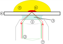
An inverted light microscope with an objective with oil immersion and a very high numerical aperture , for example 1.45, is typically used in order to be able to achieve a flat angle of incidence. Either the excitation light is coupled in at the edge of the objective so that it falls onto the cover glass at a flat angle (Cis-TIRFM, left diagram), or an additional prism is used on which an evanescent field is formed (Trans-TIRFM, right schematic drawing). In the second implementation, the demands on the objective are not so high, since such a high numerical aperture is not required.
application
TIRF microscopy can be used to examine structures that are very close (approx. 200 nm for visible light) to a surface. These can be fluorescent molecules in the membrane of a cell or close to it (see picture above). With classic fluorescence microscopy , the signal near the surface would be covered by the background scattered light. TIRF is particularly suitable for examining exocytosis .
When using a suitable camera, TIRFM is sensitive enough to detect individual fluorescent molecules. The method is therefore also used to observe individual molecules bound to a glass surface.
TIRFM can also be used in conjunction with fluorescence correlation spectroscopy (then referred to as TIR-FCS) or single-particle tracking techniques to observe and quantify the dynamics of fluorescent molecules.
literature
- NL Thompson, TP Burghardt, D. Axelrod: Measuring surface dynamics of biomolecules by total internal reflection fluorescence with photobleaching recovery or correlation spectroscopy . In: Biophys J. Band 33 , no. 3 , 1981, p. 435–454 , doi : 10.1016 / S0006-3495 (81) 84905-3 , PMC 1327440 (free full text).


