
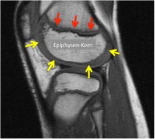
Epiphyseal plate


|

|
|
|
Skeletal scintigraphy of two children, 7 years old on the left, 15 years old on the right (not to scale). In each case intensive bone metabolism in the growth plates.
|
||

The epiphyseal plate (or growth plate, from Greek: επίφυση - "grown on", "originated") consists of a zone of strong cell division (reserve or proliferation zone) of hyaline cartilage and a transition zone (palisade zone), in which the cartilage in the longitudinal direction of the Bone hypertrophied over a short distance, dies and calcified. This zone is referred to as the opening zone in the context of the dissolution of the cartilage cells and the calcification of the cartilage base substance. The growth area is the joint between the epiphysis (end piece, support of the cartilaginous joint surface) and the metaphysis of the long bones . It is the place where the long bones grow in length due to enchondral ossification . The ossification in the epiphyseal plate is epiphyseal called. Another important place of bone growth is the secondary epiphyseal plate (physis), which encloses the bony epiphyseal nucleus. In addition, it enables the epiphyseal condyles to grow in length.
physiology
In juvenile vertebrates and humans, the long bones grow in the cartilaginous epiphyseal plate. This bone growth is called interstitial because bone cells ( osteoblasts ) migrate into the cartilage. As a result, the diaphysis becomes longer on the one hand and the epiphysis is shifted distally on the other. As soon as bone tissue completely replaces the epiphyseal plate, an individual's growth in length is complete.
There are special atlases that allow the chronological age of a child to be compared with the current skeletal age (Fig.) Due to the large number of growth plates on the hand bones, which all close at different times, there are special atlases for assessing the epiphyseal plates on the human hand skeleton , an allocation in half-year steps is possible. This means that, together with current length measurements of body size, seat size, leg length, etc., statements can be made about the expected overall growth of the child or adolescent. Due to the different gender development in girls and boys, the maturation process and thus the skeletal growth in girls is completed earlier. The first epiphyseal plates close in girls around the age of 14, the last joints in boys after the age of nineteen.
X-ray examinations of children born between 1915 and 2006 have shown that the point in time at which the growth plates close has been brought forward over the last century and thus - like puberty - occurs earlier and earlier. The reason for this is not apparent.
Diseases
In contrast to adults, in whom the entire long bone is bony except for the joint ends covered with articular cartilage , the child's bone is very vulnerable, especially in the area of the growth plate in children and adolescents. Under mechanical stress can damage give (z. B. here Apophysitis calcanei , Crohn Schlatter , X-leg ). With bacterial inflammation z. B. the upper respiratory tract can lead to bacterial abscesses in a growth plate. The capillary opening zone is a danger point for infection settlements. Severe forms of incorrect growth can occur if the diagnosis and surgical therapy with clearing out the abscess and specific antibiotic therapy is not carried out or not carried out early, which is the case above all in countries with an inadequate health system. A large proportion of global growth defects in extremities result from such traumatic or bacterial disorders in the epiphyseal plates.
Isolated injuries to the epiphyseal plate (e.g. epiphysiolysis ) and bone fractures near the joint that involve the epiphyseal plate, treated or untreated, can lead to defective growth. In the event of such injuries, it must be ensured during conservative or surgical treatment that the operation and the implants do not cause any further damage to the joints with possible incorrect growth. As with multiple epiphyseal dysplasia , growth disorders can also be hereditary.
Joint fractures of the ankle joint with the inclusion of an already partially closed epiphyseal plate are called transitional fractures . As it is multiplanar, your surgical treatment is a challenge. In addition to x-rays in 4 planes, diagnostics usually require computer tomography
See also
Individual evidence
- ^ Karl-Heinz Jungbluth, Manfred Dallek, Norbert M. Meenen. 1997. Injuries to the growth plate. The Trauma Surgeon 100: 571-586
- ^ Dietrich Starck: Embryology. A textbook on a general biological basis. 3rd, revised and expanded edition. Georg Thieme, Stuttgart 1975. There bone formation on a cartilaginous basis, p. 574f.
- ↑ Wolfgang Bargmann: Histology and microscopic anatomy of humans. 7th, revised edition. Georg Thieme, Stuttgart 1977. There chondral bone formation, pp. 131–135.
- ^ William Greulich: Radiographic atlas of skeletal development of the hand and wrist. Stanford University Press, 1999. ISBN 0804703981
- ↑ Nadja Podbregar: Children's bones mature faster today: growth plates in bones close earlier than they did 100 years ago. In: scinexx.de. December 19, 2018, accessed February 19, 2020 .