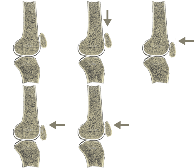
Zohlen sign

The Zohlen symbol , named after Eberhard Zohlen , appears in a common examination method in sports medicine , (accident) surgery and orthopedics . It is used to assess the retropatellar cartilage situation .
execution
With one hand the kneecap (syn. Patella) is carefully fixed and distalized (pushed towards the lower leg). By actively tensing the quadriceps , the patella is then moved cranially again. It is pressed lightly onto the femoral patellar sliding bearing by the examining hand that is holding it.
Functional anatomy
The anatomical structures involved are the back surface of the patella and the patellar slide bearing, an area of the thigh bone (syn. Femur) covered with articular cartilage , which is anatomically located within the joint capsule of the knee joint. Because of its special function, this section of the knee joint is also known as the femoropatellar joint.

The patella glides over the cranial cartilage area shown on the femur during active extension of the knee joint
The patella serves as a hypomochlion for the tendon of the large thigh extensor muscle (lat. Musculus quadriceps femoris) during extension of the knee joint. A high pressure (> 1000 N / cm 2 ) acts on your cartilage-coated rear surface and the femoral patellar sliding bearing. The main contact points are different depending on the angle at which the knee joint is bent . Active (muscular) caudalization of the patella is not possible with the knee joint extended. When the large thigh extensor muscle is tensed with the knee joint extended (isometric or to lift the leg from a lying position), the patella is therefore cranialized over a much shorter distance than when examining the Zohlen sign.
Numerous reasons ( Patelladysplasie , (Bagatell-) dreaming, patella (sub) luxations , and many others) can cause cartilage damage in the patellofemoral joint. The generic term for cartilage damage on the back surface of the patella is chondropathia patellae .
During the examination, the patella is first pushed towards the foot over the femoropatellar slide bearing while stretching the quadriceps and then pulled back from the head of the quadriceps under slight pressure from the ventral side . Different parts of the femoral patellar joint come into contact under pressure than with physiological movements.
Testimony of the test
With manual caudalization of the patella, the examiner often notices characteristic rubbing in the case of cartilage damage, which indicates irregularities in the cartilage surface, such as occur in the context of chondropathic changes.
Pain on examination may be due to cartilage damage to the back of the kneecap or an inflammatory reaction, e.g. B. by overloading the adjacent synovial membrane (peripatellar). However, the informative value is very limited, since the test is "positive even in the majority of healthy people". If the Zohlen test is negative, however, pronounced cartilage damage is rather unlikely.
Sources and individual references
- ↑ Klaus Buckup : Clinical tests on bones, joints and muscles. Thieme-Verlag, Stuttgart 1995; ISBN 3-13-100991-8