Agrin
| Agrin | ||
|---|---|---|

|
||
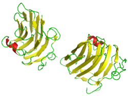
| Band model by Agrin, according to PDB 1PZ7 | ||
| Properties of human protein | ||
| Mass / length primary structure | 2,067 amino acids , 217,232 Da (protein content ) | |
| Identifier | ||
| External IDs | ||
| Orthologue | ||
| human | House mouse | |
| Entrez | 375790 | 11603 |
| Ensemble | ENSG00000188157 | ENSMUSG00000041936 |
| UniProt | O00468 | A2ASQ1 |
| Refseq (mRNA) | NM_001305275.1 | NM_021604 |
| Refseq (protein) | NP_001292204.1 | NP_067617 |
| Gene locus | Chr 1: 1.02 - 1.06 Mb | Chr 1: 156.17 - 156.17 Mb |
| PubMed search | 375790 |
11603
|
Agrin is a proteoglycan from the group of heparan sulfates . It is involved in the formation of nerve cells during embryonic development and controls the formation of contact points between nerve and muscle cells.
properties
Agrin has a molecular mass of 400 kDa and is located in the synaptic membrane of the motor endplate and other basement membranes. Its main function is the development of the motor endplate during embryonic development and the subsequent regeneration of these structures. The name comes from the ability to bind other proteins to the postsynaptic membrane and to aggregate. These proteins are primarily nicotinic acetylcholine receptors (ACh receptors), but also cholinesterases , agrin receptors and elements of the cytoskeleton and basement membrane . This enables local concentrations of the ACh receptors of up to 20,000 per square micrometer, which means that the concentration is several thousand times greater than outside synaptic areas. While Agrin ensures the accumulation of pre-existing ACh receptors, it is the protein ARIA that increases the total number of ACh receptors on the muscle surface. In humans it is encoded by the AGRN gene.
Agrin is also found in other tissues, above all in the glomerular basement membrane in the kidney and the alveolar basement membrane in the lungs.
Working mechanism
The exact mechanism of operation is not yet fully understood. One suggestion is the Agrin hypothesis by Jack McMahan: During embryonic development, Agrin is synthesized in the anterior horn of the spinal cord , transported along the axon and finally secreted into the synaptic cleft by growth cones of the growing motor neuron . This is bound by the agrin receptor in the muscle's plasma membrane, which in turn activates the MuSK protein, a tyrosine kinase, which itself is not involved in agrin binding. The MuSK protein then activates factors of an as yet incompletely understood intracellular signal cascade through autophosphorylation , which now lead, among other things, to phosphorylation and, as a result, its aggregation of the nicotinic acetylcholine receptors . Cholinesterases , sodium channels and several other proteins also aggregate in this way .
structure
Agrin consists of many different domains, which is typical of large, extracellular proteoglycans . The N terminus contains a laminin- binding domain, which ensures that the molecule is incorporated into the synaptic basement membrane after secretion by the nerve cell and remains an integral part of this structure. Furthermore , Agrin consists of many follistatin , laminin and EGF -like domains. The latter two types of domains form the C -terminal region of the molecule, which is responsible in particular for binding to the agrin receptor and thus for the formation of synaptic structures. Alternative mRNA - splicing in this part of the molecule - Three splices are described - leads to isoforms that have different activities in the construction of synaptic structures. A splice point proves to be particularly critical. Out of a total of five isoforms detected so far - theoretically a total of 16 isoforms are conceivable - two have no synaptic activity at all. In contrast to the active agrini isoforms, such inactive isoforms are also used by non-neuronal cells such as e.g. B. muscle cells produced.
Individual evidence
- ↑ Guoshan Tsen, Willi Halfter, Stephan Kröger, Gregory J. Cole: Agrin Is a Heparan Sulfate Proteoglycan . In: Journal of Biological Chemistry . tape 270 , no. 7 , February 17, 1995, ISSN 0021-9258 , p. 3392–3399 , doi : 10.1074 / jbc.270.7.3392 , PMID 7852425 ( jbc.org [accessed August 31, 2017]).
- ↑ M. Gautam, PG Noakes, L. Moscoso, F. Rupp, RH Scheller, JP Merlie, JR Sanes: Defective neuromuscular synaptogenesis in agrin-deficient mutant mice. In: Cell. Volume 85, Number 4, May 1996, pp. 525-535, PMID 8653788 .
- ↑ Kandel, Eric R .; Schwartz, James H .; Jessell, Thomas M .: Neuroscience, An Introduction . Ed .: Jessell, Thomas M. 1st edition. Spectrum, 1996, p. 111 .
- ^ Fabio Rupp, Donald G. Payan, Catherine Magill-Solc, David M. Cowan, Richard H. Scheller: Structure and expression of a rat agrin . In: Neuron . tape 6 , no. 5 , p. 811-823 , doi : 10.1016 / 0896-6273 (91) 90177-2 ( elsevier.com [accessed August 31, 2017]).
- ^ A b S. Kröger, JE Schröder: Agrin in the Developing CNS: New Roles for a Synapse Organizer. In: Physiology. 17, 2002, p. 207, doi : 10.1152 / nips.01390.2002 .
- Jump up ↑ AJ Groffen, CA Buskens, TH van Kuppevelt, JH Veerkamp, LA Monnens, LP van den Heuvel: Primary structure and high expression of human agrin in basement membranes of adult lung and kidney. In: European Journal of Biochemistry . Volume 254, Number 1, May 1998, pp. 123-128, PMID 9652404 .
- ↑ Agrinhypothese. Retrieved September 1, 2017 .
- ↑ DJ Glass, DC Bowen, TN Stitt, C. Radziejewski, J. Bruno, TE Ryan, DR Gies, S. Shah, K. Mattsson, SJ Burden, PS DiStefano, DM Valenzuela, TM DeChiara, GD Yancopoulos: Agrin acts via a MuSK receptor complex. In: Cell. Volume 85, Number 4, May 1996, pp. 513-523, PMID 8653787 .
- ^ Ad Aertsen, Adriano Aguzzi: Agrin. In: Lexicon of Neuroscience. Spektrum Akademischer Verlag, 2000, archived from the original on May 25, 2016 ; accessed on August 31, 2017 .