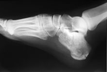
Heel bone
The heel bone (lat. Calcaneus , also Germanized calcaneus ) is a short bone of the tarsus . It is the largest and longest tarsal bone and at the same time the bone with the relatively largest proportion of bone marrow (cancellous bone).
The body of the calcaneus is roughly cuboid. Under the malleolar fork of the ankle , it is not in the middle, but offset to the side. The body runs from the back of the foot to the front - up - to the outside.
construction
At the rear end is the heel hump ( tuber calcanei ), which forms the heel ( calx ) of the foot. This is where the twin calf muscle and clod muscle come in via the Achilles tendon ( Tendo calcanei ) . Between the Achilles tendon and the heel hump is a bursa , the calcaneal tendinous bursa . On the underside of the heel cusps there are two forward-facing processes, the processus medialis tuberis calcanei and the processus lateralis tuberis calcanei . Here are the abductor hallucis , the flexor digitorum brevis and abductor digiti minimi their origins. The plantar aponeurosis also has its origin in the area of the heel hump. From processus medialis often goes heel spur from. The heel spur is a pathological and painful thorn-like ossification in the area of origin of the plantar aponeurosis.
On its underside (plantar side) are the long sole ligament and the ligamentum calcaneocuboideum plantare .
The anterior calcaneal process is located to the front . This is where the joint surface for the cuboid bone is located , which is called the facies articularis cuboidea and together with the cuboid bone forms the calcaneocuboid joint.
On the medial surface of the calcaneus there is a protrusion that "covers" the surface. This protrusion is called the sustentaculum tali . Large parts of the talus rest on this ledge. The tendon of the flexor hallucis longus muscle runs under the sustentaculum tali . This gives the muscle the effect of preventing the heel bone from buckling inwards. In addition, the tendons of the flexor digitorum longus muscle and the tibialis posterior muscle also run in this area, as do blood vessels and nerves. This space is called the tarsal tunnel .
On the upper (dorsal) side there are three articular surfaces:
- Facies articularis talaris anterior
- Facies articularis talaris media (this is located on the sustentaculum tali )
- Facies articularis talaris posterior
Located between the latter two articular surfaces of the sulcus of the calcaneus , which together with the sulcus talaris the talus one as Canalis tarsi forming tunnel designated. The facies articularis talaris anterior and the facies articularis talaris media are together part of the anterior lower ankle joint. The facies articularis talaris posterior is part of the posterior lower ankle joint.
On the side surface there is a small hump, the trochlea fibularis . This hump separates the tendons of the long and short fibula muscles .
Anatomical features of sprinters
Top sprinters often have the anatomical peculiarity of unusually short heel bones. As a result, the leverage that the Achilles tendon - and thus the calf muscles - have to cover when accelerating is significantly smaller in these athletes than in athletes with longer heel bones. The speed with which the calf muscles have to contract is therefore lower, so that higher forces are transferred to the musculoskeletal system of the foot, which ultimately enables the athlete to accelerate more quickly .
Injuries and illnesses
Heel fractures are usually caused by falls from great heights or traffic accidents, but occasionally even lower heights are sufficient. Patients cannot put weight on the heel due to pain. Often there is a bruise on the sole of the foot as well as on the inside and outside under the inner or outer ankle. A distinction is made between fractures with and without joint involvement (intra- or extra-articular fractures). Extra-articular fractures are mostly treated conservatively, while those with joint involvement and with strong displacement of the fracture fragments are often operated on and then fixed in the reduced position by means of osteosynthesis . Plates and screws are often used. The aim of the restoration is to reconstruct the geometry of the heel bone as ideally as possible. Fractures with joint involvement are often the cause of premature deformation of the lower ankle joint.
Since the heel hump serves to redirect the strength of the strong Achilles tendon to the metatarsus and forefoot, tendon irritation is frequent , especially in middle age , which can lead to achillodynia of the Achilles tendon at its heel bone or plantar fasciitis at the origin of the plantar fascia. In the event of chronic irritation, small bone noses can also form in both places, which are referred to as Haglund's exostosis on the Achilles tendon or as heel spur on the sole side of the plantar fascia.
In children during or shortly before the growth spurt, juvenile osteochondrosis , also known as Sever-Haglund's disease, can occur on the apophysis of the heel cusps due to the strong pulling of the Achilles tendon, especially during intensive sport with high running and jumping loads (such as football ) just as the Achilles tendon irritation in adults is called Haglund's syndrome, but heals painlessly and without complications when the growth is completed.
Web links
Individual evidence
-
^ Sabrina SM Lee and Stephen J. Piazza: Built for speed: musculoskeletal structure and sprinting ability. In: Journal of Experimental Biology. Volume 212, No. 22, pp. 3700-3707, doi: 10.1242 / jeb.031096
Sprinters have short heel bones . On: Wissenschaft.de from October 30, 2009.


