Colloblast
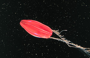
As Colloblasten or Lasso cells is called a used in prey capture cell type of the ctenophores (Ctenophora). They consist of a hemispherical head, which is anchored by a long shaft in the tentacles or their fine side threads, the tentils . The head itself is littered with numerous fine globules which, when touched, release a sticky substance so that the prey sticks to the tentacles.
Occurrence and distribution
Colloblasten found in all Tentaculata - types of comb jellies with the exception of representatives of the genus Euchlora which instead on their prey acquired stinging cells have.
They occur exclusively in the tentacles and the fine transverse threads on them, the tentils. There they form the main component of the epidermis (outer skin), in which they are surrounded by collections (clusters) of sensory and glandular cells that can be viewed as a kind of early warning and control system.
structure
Depending on the species, colloblasts have a length of 10 to 25 micrometers and a maximum diameter of 4 to 10 micrometers from the tip of the shaft to the dome-shaped head protruding from the tentillary or tentacle surface .
In the center of the head there is a star-shaped structure, the spheroidal body , from which numerous fine fibers ( fibrils ) run radially outwards. On the surface of the dome, they open into spherical bubbles with a diameter of about 1 micrometer. The thin wall of the vesicle lies directly below the cell membrane and breaks under relatively little mechanical stress, so that in this case the sticky content is released, which has a relatively high concentration of the amino acid proline , the exact composition of which is still unclear. It is assumed that the adhesive effect is based on the formation of collagen-like cross-links.
Furthermore, there are various cell organelles in the colloblast head: mitochondria , which play an important role in the energy metabolism of the cell, cisterns of the rough endoplasmic reticulum in which proteins are synthesized, Golgi complexes that organize the further processing and transport of these and other substances, and microtubules which primarily play a structural role.
The shaft consists of two externally visible components, the straight axial filament and the spirally around this slender herumgewundenen Spiralfilament formed as a hollow tube. Both are completely inside the cell, so they are surrounded by the cell membrane and connected to one another via a thin structure. Together they function as shock absorbers: violent movements emanating from prey animals in agony are cushioned in this way and cannot endanger the firm anchoring of the cell in the tentacles or tentillum.
The upper two thirds of the axial filament are taken up by the elongated cell nucleus located in the center of the shaft . At the lower end, the filament ends in the conical colloblast root, which extends into the mesoglea , the connective tissue layer below the epidermis (outer skin) .
The spiral filament arises on the spheroidal body in the center of the colloblast head and from there winds several times around the axial filament. The number of coils is species-specific and is related to the activity of the typical prey: the higher the risk that kicking prey that has got caught pulling the colloblasts out of their anchoring due to the strong mechanical stress, the more coils there are; in the copepods of the genus Euplokamis , for example, there are eleven. Finally, the spiral element opens into the colloblast root and merges there with the axial filament.
The root itself is surrounded by a multi-layered extracellular partition, the basal lamina. In the immediate vicinity there are mostly synapses of nerve cells , which has led to the previously unproven assumption that nerve impulses are involved in triggering the colloblast reaction in a manner similar to that of nettle cells.
Cell development and degeneration
Colloblasts are formed at the root of the tentacle. Auxiliary cells play a role in their differentiation and degenerate after the cell is fully developed. Their remains are often visible in electron microscopy as dark fragments on the head of the colloblast.
The spiral fragment is viewed by some scientists as a modified scourge . This is supported by the fact that its development starts from a basal body . In the embryonic stage, nine microtubules are often still present, which are arranged in the manner typical of eukaryotic flagella. According to this view, the colloblastic root corresponds in evolutionary terms to a flagellum root.
After contact with a prey animal and the activation of the adhesive bodies associated with destruction of the cell membrane, the colloblasts degenerate and must be completely replaced together with the entire tentillum or tentacle section. This replacement process takes place permanently, but can be accelerated if large tentacle sections are destroyed. Replacing an entire tentacle arm this way only takes about half a day.
Research history
Colloblasts were first identified in 1844 by the naturalist JGF Will in his publication
- Horae Tergestinae: Description and anatomy of the Akalephen observed in the autumn of 1843 near Trieste.
mentioned. The Tübingen zoologist Gertrud Benwitz described the differentiation of these cells for the first time in 1978 following electron microscopic examinations.
literature
- FW Harrison, JA Westphal: Microscopic Anatomy of Invertebrates. Volume 2, Wiley-Liss, 1991, chapter 7.
- U. Welsch, V. Storch: Introduction to cytology and histology of animals. Gustav Fischer Verlag Stuttgart 1973.
