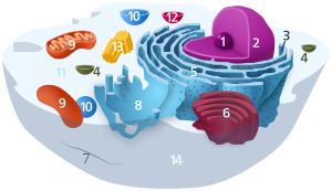
Golgi apparat

1. Nucleolus (nuclear body)
2. Cell nucleus (nucleus)
3. Ribosomes
4. Vesicle
5. Rough (granular) ER (ergastoplasm)
6. Golgi apparatus
7. Cytoskeleton
8. Smooth (agranular) ER
9 . mitochondria
10. lysosome
11. cytoplasm (with cytosol and cytoskeleton )
12. peroxisomes
13. centrioles
14 cell membrane

Transmission electron microscope image of a contrasted ultra-thin section . Cutting plane parallel to the stacking axis, the flattened, membrane-bound bags are cut transversely. There are constricted vesicles over the trans-Golgi network.

(1) nuclear membrane ,
(2) nuclear pore ,
(3) rough ER,
(4) smooth ER,
(5) ribosome on the rough ER,
(6
) transport vesicle with proteins, (7) transport vesicle ,
(8) Golgi Apparatus,
(9) cis -Golgi network,
(10) trans -Golgi network,
(11) cisterns of the Golgi apparatus.
| Parent |
| Organelle |
| Subordinate |
| Golgi membrane Golgi lumen Golgi stacks Golgi networks Golgi cisterns Protein complexes |
| Gene Ontology |
|---|
| QuickGO |
The Golgi apparatus [ ˈɡɔld͡ʒi ] (the second “g” is pronounced like the “j” in jeans ) is one of the organelles of eukaryotic cells and forms a membrane-enclosed reaction space within the cell. It is involved in secretion formation and other tasks of cell metabolism and was named after the Italian pathologist Camillo Golgi , who discovered it in 1898 during histological research on the brain.
construction
In almost all organisms, the Golgi apparatus consists of four to six - with the exception of some flagellates with up to several hundred - membrane-enclosed, mostly flat cavities, which are referred to as cisterns or dictyosomes . Usually 3 to 8, rarely up to 30, of these dictyosomes form a stack with an average diameter of 1 µm. Depending on the cell type, the Golgi apparatus can contain one to several hundred dictyosomes. The Golgi apparatus is mostly located near the cell nucleus and centrosome , which is ensured by microtubules . In some cells, however, the Golgi apparatus is not limited to this space, but is distributed throughout the cytoplasm ; this is true for most plant cells and some non-plant cells.
A clear polarization can be determined on the Golgi apparatus. The side that faces the endoplasmic reticulum (ER) and receives constricted vesicles from it, which are covered with the coat protein COP II , is called the cis-Golgi network (CGN) ; it is convex . Vesicles can also be sent from the CGN to the ER; for this purpose the vesicles are provided with a different envelope protein ( COP I ). The side facing away from the ER and closer to the plasma membrane is known as the trans-Golgi network (TGN) ; it is concave . So-called Golgi vesicles are pinched off here. The Golgi networks are several smaller cisterns and vesicles that are connected to one another.
The cisterns between the Golgi networks are called Golgi stacks , and the individual stacks contain specific enzymatic equipment . There are two models for the passage of the proteins through the Golgi apparatus, both of which are presumably applicable: On the one hand, the individual cisterns “migrate” from the cis to the trans side, while the enzymes retain them for the cistern that moves up via opposite vesicular transport (model of tank maturation). On the other hand, one observes vesicle movements through which the proteins are transported to the next cistern - in the direction of the TGN (model of vesicular transport); the Golgi apparatus is therefore a dynamic system .
If cells divide in the cell , the Golgi apparatus disintegrates and is divided between the two daughter cells, where it is then reassembled.
Structural diversity using the example of yeast
In Pichia pastoris there are usually around four Golgi stacks per cell, each made up of four cisterns. In contrast to vertebrate cells, the structure of the Golgi stacks in P. pastoris is independent of microtubules or the cell cycle. The Golgi stacks do not grow out of the nuclear envelope, but are formed on the endoplasmic reticulum (ER). Since P. pastoris has bundled structures of the ER, typical Golgi stacks arise directly next to it. In contrast, Saccharomyces cerevisiae has structures of the ER distributed throughout the cell, which form an equally scattered Golgi instead of typical Golgi stacks.
Functions
The functions of the Golgi apparatus are diverse and very complex, but can be divided into three groups according to the current state of knowledge:
- Formation and storage of secretory vesicles ( extracellular matrix , transmitters / hormones),
- Synthesis and modification of elements of the plasma membrane,
- Formation of primary lysosomes .
As already described above, the Golgi apparatus (mostly from the ER) receives vesicles that contain proteins or polypeptides; these proteins are now further modified here. Depending on the later use and the protein, different other proteins or sugar residues ( glycosylation ) of different lengths are bound to the actual protein; the structure of the protein is also changed. All these modifications take place within the Golgi apparatus, since they would lead to reactions in the cytoplasm with other cell organelles and substances, which could mean the immediate death of the cell.
If the proteins are completely modified, they are sorted in the TGN according to their destination, tied up in Golgi vesicles, provided with signal proteins (SNARE proteins) and transported to their destination via cell-internal transport mechanisms. Most proteins that are modified in the Golgi apparatus are transported out of the cell via exocytosis , so the extracellular matrix (ECM) can be modified via exocytosis , whereby it is important that all substances except the glycosaminoglycan (GAG) hyaluronan (formerly : Hyaluronic acid), which forms a significant part of the ECM, can be produced in the Golgi apparatus. The modification of the ECM contributes significantly to the intercellular communication and the stability of the tissue and is therefore one of the most important tasks of the Golgi apparatus. In addition, a cell can, for example, repair or enlarge its cell membrane; At the same time, the cell is given the opportunity to change the outer structure of the membrane, which can be beneficial to metabolism and intercellular communication.
The Golgi apparatus forms primary lysosomes . It contains lytic enzymes whose activity is optimal at a pH value of around 4.5, which means that the inside of the lysosome has to be acidified, which is ensured by proton pumps specifically built into the membrane. The interior of the lysosome is lined with proteoglycans to protect against acid . So that no wrong proteins are included when lysosomes are constricted, the lysosomal membrane is covered with mannose-6-phosphate receptors , to which the lytic enzymes, which have been modified with mannose-6-phosphates , bind.
The function of the Golgi apparatus in plant and animal cells is almost identical, but the most important task of the Golgi apparatus in plants is the production of polysaccharides for the plant cell wall ( pectins and hemicelluloses ). Since these substances have to be produced in very large quantities, this explains the enormous quantity of the Golgi apparatus in the plant cell compared to the animal cell.
Post metaphor
The Golgi apparatus basically works like the post office: It receives protein packets from the endoplasmic reticulum. Within the Golgi, these proteins are modified by removing or replacing sugar monomers. In addition, the proteins are sorted by adding identification symbols such as phosphate groups (similar to a zip code). This "post code" indicates the destination. Finally, the proteins are shipped in transport vesicles.
literature
- Bruce Alberts et al. a .: Molecular Biology of the Cell. 5th edition. Garland Science, New York 2008, ISBN 0-8153-4106-7 .
- Neil A. Campbell et al. a .: biology. 1st edition, 1st corrected reprint, Spektrum, Heidelberg 1997, ISBN 3-8274-0032-5 .
Web links
- Golgi apparatus Structure and function Text with graphics on the Golgi apparatus
- Detailed article on Camillo Golgi and his research
Individual evidence
- ↑ OW Rossanese J. Soderholm, BJ Bevis, IB Sears, J. O'Connor: Golgi structure correlates with transitional endoplasmic reticulum organization in Pichia pastoris and Saccharomyces cerevisiae . In: The Journal of Cell Biology . tape 145 , no. 1 , April 5, 1999, ISSN 0021-9525 , p. 69-81 , PMID 10189369 , PMC 2148216 (free full text).
- ↑ Reece, Jane B.,: Biology: a global approach . Tenth ed., Global ed. Boston, ISBN 978-1-292-00865-3 .