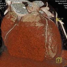
Computed tomography of the heart
The computed tomography of the heart (cardiac CT) is a special CT angiography of the coronary arteries .
Indications
A clinical benefit was shown in individual patient studies for the following indications :
- The suspicion of coronary artery disease (CHD) with medium clinical probability.
- For monitoring the progress of a coronary artery bypass .
- For the evaluation of heart valve diseases.
- For the evaluation of cardiac masses.
Cardiac CT has a good negative predictive value . The examination is therefore well suited to rule out coronary heart disease. However, the examination is poorly suited to prove coronary heart disease.
Contraindications
A pregnancy is an absolute contraindication . As contrast media containing iodine is used, a contrast agent allergy, hyperthyroidism or impaired kidney function are relative contraindications. Cardiac arrhythmias , coronary artery stents, or tachycardia usually reduce the image quality and make CT coronary angiography less useful.
execution
Lime score
Before the actual CT angiography of the coronary arteries, a so-called Kalk-Score examination is often carried out. The recordings are made without the use of contrast medium. While the slice thickness for CT coronary angiography is in the range of approx. 1 mm, it is between 3 and 5 mm for Kalk-Score recordings. The amount of coronary calcium is measured individually and quantitatively for each vessel. The extent to which the amount of calcium has a prognostic value on the course of coronary heart disease is controversial. A subsequent CT coronary angiography should be avoided in the case of high amounts of calcium, since the evaluation of calcified vessels is imprecise or even impossible.
CT angiography of the heart
Due to the small diameter of the coronary vessels and the movement of the heart, a CT machine with a high spatial and temporal resolution must be used for the examination. Since the detector width of almost all CTs (as of 2011) is smaller than the diameter of the heart, the image has to be composed of recordings from several cardiac cycles. The wider the detector of the CT used, the fewer cardiac cycles are required for a recording (approx. 5 to 10 heartbeats for a 64-line CT) and the lower the probability that image artifacts will be generated by a cardiac arrhythmia. In order to achieve a lower heart rate and variability, a beta blocker is administered if possible before the examination . Glycerol trinitrate is also often administered to enlarge the diameter of the coronary vessels. An EKG is recorded in parallel to the examination . Iodine-containing contrast medium is applied to visualize the vessels . Stenoses within the coronary vessels can be recognized and measured. A distinction can be made as to whether the stenosis is caused by calcified plaques or by non-calcified plaques. If coronary heart disease (CHD) is suspected on CT , invasive cardiac catheterization is usually required for confirmation . Patients at high risk of CHD should therefore be examined primarily with a cardiac catheter in order to avoid double exposure. In patients with a low to medium probability of CHD, a cardiac CT examination appears to be suitable to rule out CHD.
Radiation exposure
The radiation exposure from a cardiac CT is between 5 and 30 mSv, depending on the CT device used and the examination protocol, and 1 to 3 mSv, according to more recent information. This corresponds to 1 to 15 times the natural radiation exposure per year in Germany. The risk of radiation cancer is difficult to estimate and, according to the calculation formula of the International Commission on Radiological Protection, lies between 1.5: 1,000 and 2.5: 10,000 additional tumor diseases per examination. By using modern CT scanners with appropriate examination protocols and reconstruction algorithms, it is possible in individual cases to carry out a CT coronary angiography with approx. 1 mSv or even in the sub-millisized range; the average radiation exposure is below 10 mSv.
Alternative investigation procedures
The gold standard for the representation of the coronary vessels is the examination by means of a cardiac catheter / coronary angiography . In special cases, CT angiography of the heart can be an alternative examination method to cardiac catheters.
The EKG enables the diagnosis of excitation propagation disorders in the heart, such as B. occur in a recent or old heart attack. The EKG is also suitable for identifying arrhythmias.
With echocardiography movement disorders, blood clots and valve leaks can be determined.
The myocardial scintigraphy is associated with radiation exposure investigation which acute and chronic circulatory disorders can present in the heart muscle.
The cardiac MRI examination is a procedure without radiation exposure that can depict wall movement disorders and functional disorders. However, it has not yet been able to establish itself as a standard procedure.
literature
- Dewey: Coronary CT Angiography . Springer Verlag, 2008, ISBN 3-540-79843-9
Web links
Individual evidence
- ↑ Guidelines for the use of computed tomography in the diagnosis of the heart and the great thoracic vessels. (PDF; 31 kB) (No longer available online.) In: AG Herzdiagnostik der Deutschen Röntgengesellschafte.V. 2009, archived from the original on October 14, 2009 ; Retrieved September 12, 2010 .
- ↑ DE Winchester, DC Wymer, RY Shifrin, SM Kraft, JA Hill: Responsible use of computed tomography in the evaluation of coronary artery disease and chest pain. In: Mayo Clinic proceedings. Volume 85, number 4, April 2010, pp. 358-364, doi: 10.4065 / mcp.2009.0652 , PMID 20360294 , PMC 2848424 (free full text) (review).
- ↑ Positive calcium score - risk factor or expensive wrong track? In: Doctors newspaper. March 31, 2005, accessed September 12, 2010 .
- ↑ Jürgen Freyschmidt : manual diagnostic radiology. Cardiovascular system Springer Verlag , 2007, ISBN 3-540-41420-7 , chapter 1.4.1.1
- ↑ Estimated Radiation Dose Associated With Cardiac CT Angiography. In: The Journal Of the American Medical Association. 2009, accessed September 12, 2010 (English, 301 (5): 500-507.).
- ↑ Gerd Herold : Internal Medicine 2016
- ^ Dose in x-ray computed tomography. In: Physics in Medicine and Biology. February 7, 2014, accessed January 30, 2014 (English, 59 (3): R129-50).
