Port catheter
The port catheter or port system (from Latin porta , "gate") is a permanent subcutaneous access to the venous blood circulation or, in rare cases, to the abdominal cavity. Port catheters are also used in long-term epidural anesthesia with particularly thin catheters and a filter .
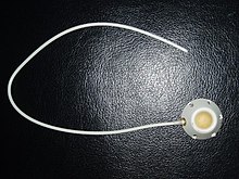
A port catheter consists of a chamber called a port, which is covered with a thick silicone membrane , and an attached tube that serves as a central venous catheter and is passed through a vein so that its open end lies just in front of the right atrium of the heart comes. The chamber can either consist of titanium , stainless steel , ceramic , plastic or a composite of the aforementioned materials. The port catheter is implanted as part of a surgical procedure.
In the main application, access to the bloodstream is established by percutaneous piercing with a cannula through the silicone membrane into the chamber . Via the cannula opening in the port chamber, a drug or a preparation can now be added to the blood stream by infusion or blood can also be withdrawn, taking into account the special features of a port catheter.
application areas
A port catheter is primarily used in the therapy of oncological diseases and in the treatment of diseases for which a frequent and safe venous or arterial access is required; Furthermore, if, due to anatomical or physiological conditions or a known or expected pharmacological effect, the use of peripheral vascular accesses for the administration of liquid medicaments, medicament preparations or therapeutics does not appear or is not possible. A port catheter can also be used to draw blood and to administer blood and blood products.
The port, which is implanted under the skin, is protected from external influences and enables the patient wearing it to maintain the freedom of movement that has been used until now. Port catheters thus enable a high quality of life. In times when there is no therapy, the patients can continue to do their usual daily activities, including showering, bathing and swimming. Even diving is possible without restrictions as far as the port catheter is concerned.
Useful life
The dwell time and therefore the period of use can be up to five years and longer. There have already been reports of patients who had "forgotten" their port over time and which was still functional and functional after more than 10 years. Nevertheless, for periods longer than five years, further use / re-use must be clarified with the responsible doctor, taking into account the medical indication and the medications administered via the port in the past and to be administered in the future.
There is nothing wrong with removing a port system once the therapy has been completed. The procedure is similar to that of port implantation. The time for this should also be discussed with the responsible doctor.
Implantation / application
The procedure is performed under local or general anesthesia under sterile conditions.
In principle, all larger veins, through which a central venous catheter can be placed, can be used as an access route for the port catheter.
- The most frequently used surgical technique involves dissection (sectio vein ) of the cephalic vein . A small skin incision is made in the so-called sulcus deltoideopectoralis , i.e. the area of the transition from the deltoid muscle to the large pectoral muscle on the front of the chest wall. From this incision, the cephalic vein is opened with a small incision and the catheter is inserted. A little apart from this, the port chamber is placed in a small pocket in the subcutaneous fatty tissue on the pectoral muscle and thus the first or second rib.
- It is also possible initially without cutting, for example, the internal jugular or the subclavian vein in Seldinger technique punctured and the catheter inserted into the vein. As described above, the port chamber is placed in the subcutaneous fatty tissue via a small incision away from the puncture site and, starting from the puncture site, the catheter is pulled through the subcutaneous tissue to the skin pocket (tunneled). This tunneling also serves as a later natural infection barrier.
A radiological check of the catheter's position is carried out in all procedures, including for documentation. Then the catheter is shortened to the required length outside the vein up to the final position of the port chamber in the skin pocket and connected to the port chamber. In the next step, the port chamber in the skin pocket is sewn to the fascia below . Then the skin incision is surgically closed (sutured). With the rib as an "abutment", the port can now be punctured ("pierced").
As mentioned at the beginning, there are other types of port catheter access for special applications, such as B. via the arteria hepatic , peritoneal or epidural, which, however, will not be explained further here. The basic function of the port always remains the same: primarily the repeated administration of drugs or preparations over a longer period of time.
Puncture
The puncture of the port (port puncture) is a medical act that can be delegated to nursing staff in order to repeatedly administer medication or infusion solutions according to an individual schedule in accordance with the medical prescription. In fact, it can be a port catheter puncture or a port needle change. Special port needles are always used for the puncture ( Huber needle , Gripper needle ), which, unlike normal injection needles - due to the special shape of their cannula tip - cannot punch out any particles from the silicone membrane of the port, which would render the port unusable and ensure that it is After removing the needle, the silicone membrane is fully closed again and no medication can escape into the tissue.
Action:
- The port is not punctured when the patient is lying down, but in the "Beach Chair Position" ( German "sun lounger position" - sitting at an angle of approx. 60 ° to almost upright), the tissue masses in the upper body follow gravity so well and are "in the correct position before the puncture "
- a pressure-stable support of the patient is created in the back in order to minimize recoil during the puncture
- sterile work
- the exposure time of the disinfectant used must be observed
- A circular wipe disinfection of the skin area above the port is carried out from the inside out
- the materials used must be kept sterile
- No overpressure may be built up via the port needle
- Therefore, no syringes smaller than 10 ml are used for this (the smaller the syringe plunger, the higher the pressure generated with the same force)
- Compliance with the five times R rule (drug safety)
- The fixation of the port needle should be transparent so that the puncture site of the needle is visible. If there is reddening or even pustules at the puncture site, the needle must be withdrawn.
- The port catheter must be flushed with sufficient saline solution before the needle is removed. For this purpose, the port catheter is rinsed with at least 2 × 20 ml of saline solution via the port immediately after a blood sample has been taken. Rinsing and blocking with the addition of heparin after applications via port catheters is still controversial in Germany because there are no guidelines on this to date.
Before and after applications, port catheters are usually flushed with a saline solution. The liquid instilled before a break in therapy, which then remains in the system as a static column of liquid until the next use, is called a so-called block solution, regardless of its chemical composition. Adding heparin to this rinsing and blocking solution, which is supposed to prevent possible thrombosis of the catheter contents during therapy breaks, has no advantages, which has meanwhile been proven. In addition, there is no blood in a port catheter that has been professionally rinsed and blocked with saline solution, but a static column of liquid from this previously applied saline solution, which makes the addition of heparin appear irrational on closer inspection.
Also, due to its pharmacokinetics, heparin could not prevent possible thrombosis, but only delay it. The half-life of heparin (UFH) is approximately 1.5 to 2 hours. With a port catheter block, periods from days to several weeks or even months have to be bridged. On top of that, heparins consist of polysaccharides, i.e. sugars. Sugar promotes germ growth. Instilling solutions based on recommendations into port catheters, which should remain in it for a longer period of time as a static column of liquid, but unfavorably at the same time support bacterial growth, is a dilemma to be discussed in detail.
The pain in the surgical area that lasts for a few days is treated with analgesics and anti-inflammatory drugs .
Complications
Possible complications can be infections , hemorrhages, pneumothorax , hemothorax or thrombosis . The pinch-off , widely described in the specialist literature, is the breakage of the catheter at the level of the collarbone with the point of intersection with the first rib, if the catheter's venous access was made directly there or medially. If the catheter is even separated, the catheter fragment remaining in the vascular system can migrate further in the direction of the blood flow. The cause is material fatigue of the catheter due to repeated bruising as a result of (unavoidable) physical activity involving the shoulder girdle. To prevent pinch-off , a more lateral puncture is recommended or a different technique is to be used (e.g. venous access after dissecting the cephalic vein or access via the jugular vein).
Catheters have also been reported to run poorly or become completely blocked. Deposits in the port chamber or on the inner wall of the catheter, especially after many infusions with nutritional solutions, can be a cause. Ports with modern, flow-optimized chamber geometry seem to have advantages here. When the needle is pulled out of the port, the resulting change in volume also draws in a small amount of blood at the end of the catheter. This can coagulate and lead to a partial or complete catheter occlusion. This can be prevented with a suitable technique in which a little rinsing solution is pressed in with the syringe while the needle is being withdrawn. However, if port catheters are no longer patency, medical action is indicated to restore patency. Rinsing attempts by means of (over) pressure are not indicated in any case.
Manufacturing
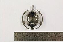
The illustration opposite shows a port made from a titanium-aluminum alloy. This essentially consists of three components:
- the chamber with the base plate underneath, which were turned in one piece from a solid blank,
- the inserted catheter connection that is fixed by the laser welding manufacturing process
- and a shrunk-on top
The sectional view shows a section through the diameter of the component, which illustrates the bond between the base plate and the chamber and the dividing line between the chamber and the upper part. The silicone membrane, through which drugs can be administered via a cannula, lies on this upper part. At the widest point (the base plate) the port has a nominal diameter of around three centimeters.
The structure of the chamber with the base plate underneath is very fine and evenly arranged; it indicates that no deformation has taken place. Instead, the blank is turned into its final shape. Under the microscope, grooves can be seen that run on the surface of the base plate and the chamber.
In order to establish a connection between the catheter and the port, a catheter connector must be attached to the chamber. To do this, a hole is first drilled in the chamber of the port; the drill is continuously cooled in order to prevent a change in the structure of the titanium. In the next step, the catheter connector is inserted into the prepared borehole and positioned. In order to create an irreversible connection between the catheter and the port, a circular weld seam is placed around the neck of the catheter connector. Since titanium has a high affinity for oxygen, the weld seam is created in a vacuum using a high-precision laser. This also prevents the structure of the titanium from changing adversely.
The upper part, to which the silicone membrane is attached, is made from the solid by prior turning. The upper part and the chamber are joined by means of shrinking. As can be seen in the adjacent picture, there are circumferential grooves on the chamber and the upper part in order to prevent the joining partners from being removed. First, the upper part is heated so that the material expands and is placed on the chamber. A firm bond is formed when it cools down.
literature
- A. Surov, K. Jordan, M. Buerke et al: Port Catheter Insufficiency: Incidence and Clinical-Radiological Correlations. Onkologie 31 (2008), pp. 455-461.
- Ulf K. Teichgräber, Robert Pfitzmann, Herbert AF Hofmann: Port systems as an integral part of chemotherapy . In: Dtsch Arztebl Int . No. 108 (9) , 2011, pp. 147-154 ( abstract ).
- H. Haindl, H. Müller, E. Schmoll (Eds.): Port catheter systems. Practical information on indications, implantation technology, handling. Springer, 1993, ISBN 3-540-56316-4 .
- B. Stevens, SE Barton, M. Brechbill et al: A Randomized, Prospective Trial of Conventional Vascular Ports vs. the Vortex "Clear Flow" Reservoir Port in Adult Oncology Patients. Journal of Vascular Access Devices 2 (2000), pp. 37-40. (Abstract)
- GA Goossens et al .: Comparing normal saline versus diluted heparin to lock non-valved totally implantable venous access devices in cancer patients; a randomized, non-inferiority, open trial. Annals of Oncology 24 (2013), pp. 1892-1899.
- Lutz Steinmüller, Marc Olaf Liedke and Margret Liehn: Shunt and Port Systems , in: Margret Liehn, Brigitte Lengersdorf, Lutz Steinmüller and Rüdiger Döhler : OP manual. Basics, instruments, operating procedures , 6th, updated and expanded edition. Springer, Berlin Heidelberg New York 2016, ISBN 978-3-662-49280-2 , pp. 321–327.
Individual evidence


