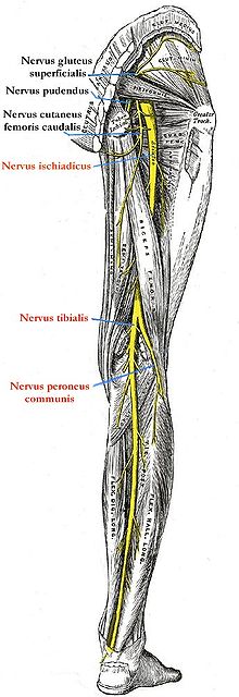
Sciatic nerve
The sciatic nerve (pronounced [ 'ɪʃias ] or [ ' ɪsçias ]), also known as the sciatic nerve (also called " sciatic " , "hip nerve" or " ischial nerve " ; os ischii = "ischium"), is a peripheral nerve of the lumbosacral plexus (lumbar nerve ) Cross braid). It is the strongest nerve in the body. In humans it has its origin in the last lumbar and the first three sacral spinal cord segments (4th lumbar segment to 3rd cross segment of the spinal cord , L4-S3). In domestic mammals, it arises from L6 – S2 as a direct continuation of the so-called lumbosacral trunk .
course
The sciatic nerve pulls over the major ischiadic incisura or the majus ischiadic foramen (more precisely through the infrapiriforme foramen ) to the extensor side of the hip joint and then on the back of the thigh , covered by the knee flexors ( ischiocrural muscles ) in the direction of the hollow of the knee . On its way, it sends branches, which vary somewhat in the individual mammals, for motor innervation to some thigh muscles:
- Musculi gemelli ,
- Quadratus femoris muscle ,
- Obturator internus muscle ,
- Biceps femoris muscle ,
- Semitendinosus muscle as well
- Semimembranosus muscle .
On the thigh, the sciatic nerve divides into:
- Nervus fibularis communis (also Nervus peroneus communis) and
- Tibial nerve .
In some mammals, the above muscle branches only start from these branches.
On closer inspection, the sciatic nerve does not actually exist (at least in humans), but is a relic from the past of anatomical-morphological research. The common fibular nerve and the tibial nerve emerge separately from the sacral plexus . Shortly before they pass through the infrapiriforme foramen , in most people they are surrounded by a thin layer of connective tissue, from which they emerge at the hollow of the knee at the latest (deep division). In some people the nerves emerge from the connective tissue sheath earlier (high division) and in still others it does not exist at all. There is no exchange of nerve fibers within this connective tissue sheath, so that all flexors (with the exception of the short head of the biceps femoris muscle ) of the knee and upper ankle joint are supplied by the tibial nerve and the extensors and pronators of the ankle joints by the common fibular nerve. The sciatic nerve is therefore “only connective tissue”. This can easily be demonstrated with dissections by splitting the connective tissue sheath (sciatic nerve) from the hollow of the knee upwards with the finger without any effort. This is not possible with "real" nerves . The situation is similar, for example, with the vestibulocochlear nerve .
Diseases
Sciatic nerve paralysis is often associated with pelvic fractures, thigh fractures, or dislocations of the sacrum and iliac joint . In small animals, injury can occur from an intramuscular injection into the hamstring muscles . If the sciatic nerve is damaged, there is no flexion in the knee joint when the flexor reflex is triggered .
A neuralgia of the sciatic nerve is usually briefly referred to as sciatica (actually: sciatica) and was first described by Domenico Cotugno (1736–1822). A characteristic pain occurs when the nerve is stretched (extended knee, bent hip) . In the so-called Lasègue test , the patient lies on their back with their legs straight, but feet in a neutral position. Passively lifting one leg causes pain on the diseased side in sciatica problems.
literature
- Martin Trepel: Neuroanatomy. Structure and function. 3. Edition. Urban & Fischer, Munich et al. 2004, ISBN 3-437-41297-3 .
- Franz-Viktor Salomon: nervous system, systema nervosum. In: Franz-Viktor Salomon, Hans Geyer, Uwe Gille (Ed.): Anatomy for veterinary medicine. Enke, Stuttgart 2004, ISBN 3-8304-1007-7 , pp. 464-577.
