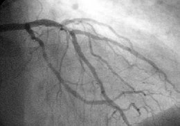
Coronary angiography

The coronary angiography is an angiography of the coronary arteries and thus a special form of x-ray examination , in which the coronary arteries are displayed. It is carried out as part of a cardiac catheter examination . X-rays make an X-ray contrast medium , which is injected into the lumina of the coronary arteries via a cardiac catheter, visible. The recordings were previously documented on film or video material, nowadays on digital storage media .
indication
It is used to diagnose the shape of the coronary vessels (coronary arteries) and to localize stenoses and their type and extent. Coronary stenoses can be expanded with a balloon catheter in the same session ( percutaneous transluminal coronary angioplasty , PTCA) and kept permanently open with the help of a stent . By filling the left heart with contrast agent, disturbances of the contraction process of the heart can be made visible under fluoroscopy (levocardiography).
Diagnostic coronary angiography is indicated in patients
- who have developed acute coronary syndrome
- who have persistent, higher-grade angina pectoris ( CCS class III and IV) during drug therapy
- with pathological results of the non-invasive examinations, regardless of the severity of the angina pectoris
- who have survived sudden cardiac arrest or life-threatening irregular heartbeat (ventricular arrhythmia)
- with symptoms of chronic heart failure .
evaluation
The narrowing of the vessel diameter (degree of stenosis), the morphology of the constriction and the still existing flow are used to describe a constriction.
Degree of stenosis
The extent of a stenosis is given as a percentage of the lumen no longer perfused. Here the longitudinal diameter is given , not the cross-section. A narrowing of the diameter of 75% corresponds to the narrowing of the area of the cross-section to 90%, ie only 10% of the remaining lumen is flowed through. As recommended by the American Heart Association , the following classifications are commonly used.
| Degree | Stenosis | description | |
|---|---|---|---|
| diameter | Cross-section (area) | ||
| 0 | <25% | <44% | Contour irregularities, diffuse coronary sclerosis |
| I. | 25-50% | 45-75% | minor or subcritical stenosis |
| II | 50-75% | 75-94% | moderate stenosis |
| III | 75 - 90% and> 90% | 94 - 99% and> 99% | severe or critical stenosis |
| IV | 100% | complete closure | |
Stenosis morphology
According to the recommendation of the ACC / AHA , the following classification is used to describe bottlenecks:
| Stenosis type | description |
|---|---|
| A. | concentric, short (≤ 1 cm), easily accessible, straight vascular segment, regular contour, little calcified |
| B. | Eccentric, elongated or tubular (1–2 cm), moderately angled (45–90 °), in the area of an ostium , a side branch or a branching of vessels (bifurcation), moderately to severely calcified, thrombus . B1 = 1 criterion met; B2 = 2 or more criteria met |
| C. | diffuse lesion, elongated (> 2 cm), pronounced tortuous vessel, extremely angled segment (> 90 °), no significant side branch protected, degenerated venous bypass C1 = 1 criterion met; C2 = 2 or more criteria met |
Coronary flow (TIMI classification)
According to the thrombolysis in myocardial infarction classification (TIMI), the coronary flow in the vicinity of a vascular occlusion or a narrowing (stenosis) is described semiquantitatively.
| TIMI river | description |
|---|---|
| 0 | no antegrade flow distal to the occlusion |
| 1 | Contrast agent can be displayed distally, but does not fill the entire vascular bed |
| 2 | Contrast medium fills the entire vessel distally, but the inflow and outflow are delayed |
| 3 | normal flow |
Risks and Complications
Main article: Risks and complications of cardiac catheterization
Overall, the risks of coronary angiography can be described as moderate. An increase in risk arises from obesity , impaired kidney function, generalized arteriosclerosis, etc. The most common, but in most cases easily manageable complication is a bleeding into the puncture site of the groin. Furthermore, problems can arise due to the iodine-containing contrast agent used (kidney damage, allergic reaction, derailment of an overactive thyroid , temporary neurological problems). In rare cases, it can also cause cracks of the coronary vessels with life-threatening bleeding into the pericardium ( pericardial tamponade ) and come to infarctions. Also strokes occur in very rare cases.
literature
- Announcements: National Care Guideline for Chronic CHD (clinically relevant excerpts from the guideline) . Dtsch Arztebl 2006; 103 (44): A-2968 / B-2584 / C-2484
Web links
- Information about coronary angiography with some film examples ( Memento from October 21, 2004 in the Internet Archive )
Individual evidence
- ↑ MA Pantaleo, A. Mandrioli et al: Development of coronary artery stenosis in a patient with metastatic renal cell carcinoma treated with sorafenib. (PDF; 287 kB) In: BMC Cancer. 2012, 12: 231 doi: 10.1186 / 1471-2407-12-231 ( Open Access )
- ↑ a b Krakau, I. / Lapp, H. (Ed.): The heart catheter book - Diagnostic and interventional catheter technology . 2nd Edition. Thieme, Stuttgart, New York 2005, ISBN 3-13-112412-1 , pp. 171-173 .
- ^ Harrison (ed.): Internal medicine . 16th edition. 2005.
- ↑ a b Mewis, Riessen, Spyridopoulos (Ed.): Cardiology compact - Everything for ward and specialist examination . 2nd Edition. Thieme, Stuttgart, New York 2006, ISBN 3-13-130742-0 , pp. 116-117 .

