Liposarcoma
| Classification according to ICD-10 | |
|---|---|
| C49.- | Malignant growth of other connective tissue and other soft tissue |
| ICD-10 online (WHO version 2019) | |
| Classification according to ICD-O-3 | |
|---|---|
| 8850/3 | Liposarcoma NOS |
| 8851/3 | Well differentiated liposarcoma |
| 8852/3 | Myxoid liposarcoma |
| 8853/3 | Round cell liposarcoma |
| 8854/3 | Pleomorphic liposarcoma |
| 8855/3 | Mixed cell liposarcoma |
| ICD-O-3 first revision online | |

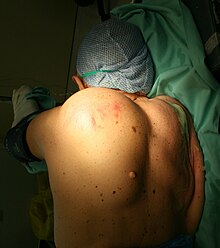
The liposarcoma is a rare malignant tumor of the soft tissue ( sarcoma ), which has the fine-tissue characteristics of fat cells or fat cell precursors. With a share of 16–18%, liposarcoma is the second most common soft tissue sarcoma after malignant fibrous histiocytoma . The first description of liposarcoma as a disease entity was in 1857 by Rudolf Virchow .
Epidemiology
The international incidence of liposarcoma is given as around 2.5 new cases per million population per year. With a mean age of onset of 50 years, it is an adult tumor that is rarely observed in children and young adults. Men are affected slightly more often than women. Geographical or ethnic frequency differences have not yet been reported.
etiology
The underlying causes of the development of a liposarcoma are largely unexplained. A possible relationship to previous injuries and exposure to ionizing radiation is described. The lipoma , a benign and much more common fatty tissue tumor, is not a typical precursor to liposarcoma, but according to some authors it should be able to form its starting point in individual cases. Other sources dispute this view and point out that the transition from lipoma to liposarcoma has never been convincingly documented.
pathology
From a macroscopic point of view, liposarcomas are often relatively good and often even capsule-like, nodular or lobed, yellowish to gray-white tumors which, depending on their location, can reach a considerable size and weight of several kilograms. The apparently good delimitation can prove to be deceptive insofar as smaller tumor settlements are sometimes found in the vicinity of the main tumor. Liposarcomas are found primarily in the deep soft tissue of the lower extremities (59%), the upper extremities (16%), the retroperitoneum (15%) and the trunk (8%). The thighs are particularly often affected (41%).
The histological (i.e. histological) examination allows a distinction to be made between several subtypes of liposarcoma, which show a different prognosis and which in some cases also occur preferentially in certain body regions:
| Histological subtype | Relative frequency | Dedifferentiation | photos |
|---|---|---|---|
| Well differentiated liposarcoma | 40-45% | low grade | Macroscopy |
| Myxoid / round cell liposarcoma | 30-35% | moderate / high | Macroscopy histology |
| Pleomorphic liposarcoma | 5% | highly | Macroscopy |
| Dedifferentiated liposarcoma | Rare | highly | Macroscopy |
The degree of dedifferentiation of a liposarcoma indicates how much the tumor tissue differs morphologically from the mature adipose tissue. This is important because with increasing tissue immaturity, an increasingly malignant biological behavior of the tumor and a poorer prognosis are to be expected (aggressive local growth, tendency to relapse , metastasis ). The terms atypical lipomatous tumor or atypical lipoma are sometimes used for well-differentiated liposarcomas , since on the one hand they can be morphologically very similar to a lipoma and, moreover, do not metastasize in the absence of tumor progression, so that they lack a prognostically significant feature of malignant tumors. It is now generally accepted that round-cell liposarcomas represent a dedifferentiated (tissue immature and thus more malignant behavior) variant of myxoid liposarcoma .
Molecular pathology
Genetic changes are common, affecting, among other things a region on the long arm of chromosome 12 (12q13-15) with amplification of the MDM2 gene ( murine double minute oncogene ) and the cyclin-dependent kinase 4 encoding gene CDK4. The associated overexpression of the corresponding genes can be detected at the RNA and protein level and, under certain circumstances, contribute to the differentiation from benign lipomas as well as from other soft tissue sarcomas .
Clinical symptoms
Liposarcomas often only become clinically noticeable in more advanced stages as deep-lying, slowly growing tumorous tissue. The exact symptoms are mainly determined by the location of the tumor. General symptoms that may be associated with tumor growth include tiredness, fatigue, weight loss, nausea and vomiting.
diagnosis
Imaging methods such as computed tomography , magnetic resonance tomography , angiography or scintigraphy provide diagnostic information and enable an assessment of the spread of the tumor. A biopsy and a histological examination of the tumor tissue obtained by a pathologist are usually required for definitive confirmation of the diagnosis .
therapy
The most promising therapeutic approach is the complete surgical removal of the tumor while maintaining a sufficient safety margin. Other therapy options are local radiation and chemotherapy . Although liposarcoma is considered to be the most radiation-sensitive sarcoma, an increase in survival time through radiotherapy has so far not been convincingly shown in scientific studies. Chemotherapy for liposarcoma is currently still of an experimental nature.
forecast
In addition to the possibility of a complete surgical removal, the chances of healing depend on which subtype of liposarcoma is present. The well-differentiated and most myxoid liposarcomas show a favorable prognosis with a five-year survival rate of 100 and 88 percent, respectively. One of the reasons for this is that these forms have little tendency to form metastases . By contrast, around 50 percent of patients with round-cell or poorly differentiated liposarcoma die from their tumor disease within five years. Metastatic tumor settlements mainly affect the lungs (20%), bones (8%), lymph nodes (6%) and the liver (5%).
literature
- AN Khan et al: Liposarcoma, Soft Tissue. (March 12, 2008);
- RA Schwartz et al .: Liposarcoma. (April 18, 2008);
Individual evidence
- ↑ FM Enzinger and SW Weiss: Soft tissue tumors. 2nd Edition, St Louis, MO: Mosby-Year Book, 1998, pp. 346-382.
- ↑ R. Virchow: A case of malignant fatty tumors partly in the form of a neuroma. In: Virchows Arch A Pathol Anat Histopathol 11, 1857, pp. 281-288.
- ^ Z. Ahmed et al.: Pleomorphic liposarcoma in a ten year old child. In: J Pak Med Assoc 54, 2004, pp. 533-534. PMID 15552292
- ↑ E. Vocks et al: Myxoid liposarcoma in a 12-year-old girl. In: Pediatric Dermatology 17, 2000, pp. 129-132. PMID 10792803
- ↑ RA Schwartz, among others: Liposarcoma: Overview. 16 July 2009
- ↑ SD Newlands et al .: Mixed myxoid / round cell liposarcoma of the scalp. In: Am J Otolaryngol 24, 2003, pp. 121-127. PMID 12649828 (Review)
- ^ Nishimoto et al .: A rare case of burn scar malignancy. In: Burns 22, 1996, pp. 497-499. PMID 8884015
- ↑ H. Ninomiya include: Post Radiation sarcoma of the chest wall: report of two cases. In: Surg Today 36, 2006, pp. 1101-1104. PMID 17123140
- ↑ D. Demir et al .: Radiation-induced liposarcoma of the retropharyngeal space. In: Otolaryngol Head Neck Surg 134, 2006, pp. 1060-1062. PMID 16730558
- ↑ Z. Orosz et al .: Pleomorphic liposarcoma of a young woman following radiotherapy for epithelioid sarcoma. (PDF; 190 kB) In: Pathol Oncol Res 6, 2000, pp. 287-291. PMID 11173662
- ↑ T. A Nickloes include: lipomas. 16 March 2010
- ↑ a b R. D. Brasfield and TK Das Gupta: Liposarcoma. In: CA Cancer J Clin 20, 1970, pp. 3-8. PMID 5005753
- ^ W. Remmele et al.: Pathology (head and neck region, soft tissue tumors, skin) 3rd edition, Verlag Springer, 2008. Excerpt from the Google book search
- ↑ MB Binh et al: MDM2 and CDK4 immunostainings are useful adjuncts in diagnosing well-differentiated and dedifferentiated liposarcoma subtypes: a comparative analysis of 559 soft tissue neoplasms with genetic data. In: Am J Surg Pathol 29, 2005, pp. 1340-1347. PMID 16160477
- ↑ N. Sirvent et al .: Detection of MDM2-CDK4 amplification by fluorescence in situ hybridization in 200 paraffin-embedded tumor samples: utility in diagnosing adipocytic lesions and comparison with immunohistochemistry and real-time PCR. In: Am J Surg Pathol 31, 2007, pp. 1476-1489. PMID 17895748
- ^ RL Jones et al .: Differential sensitivity of liposarcoma subtypes to chemotherapy. In: Eur J Cancer 41, 2005, pp. 2853-2860. PMID 16289617
- ↑ RA Schwartz, among others: Liposarcoma: Treatment and Medication. April 18, 2008
- ↑ RA Schwartz, among others: Liposarcoma: follow-up. April 18, 2008
Web links
- Liposarcoma (English)
- Histopathology India: Liposarcoma (English)


