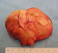
Lipoma
| Classification according to ICD-10 | |
|---|---|
| D17 | Benign new formation of adipose tissue |
| D17.9 | Lipoma |
| ICD-10 online (WHO version 2019) | |
A lipoma , also known as a benign fat tumor , is a benign tumor of the adipose tissue cells ( adipocytes ).
Incidence and distribution
With a share of around 16 percent, lipomas are the most common mesenchymal tumors in humans. The prevalence of lipoma in the soft tissues is estimated at 2.1 per 100 people. Lipomas are much more common than the related malignant liposarcomas . The ratio is around 100: 1.
At 15 to 20 percent, they are most common in the head and neck region. The shoulder and back are also very often affected. On the other hand, they are very rarely found in the fingers , for example ; there are fewer than 20 cases described in the literature.
The lipomas of the soft tissue can be divided into superficial and deeper lying. The superficial lipomas occur subcutaneously and make up 16 to 50 percent of all soft tissue tumors. They usually occur in the fifth to seventh decades of life. Deep-seated lipomas are much rarer than superficial ones, with a share of 1 to 2 percent. Since deep-seated lipomas are rarely clinically relevant and are usually only an incidental finding of a radiological examination, some authors assume a significantly higher prevalence. Deep-seated lipomas in the extremities are usually intra- or intermuscular. They are also often called infiltrating lipomas . This type of lipoma occurs mostly in patients between the ages of 30 and 60 years in the lower extremities (45 percent), trunk (17 percent), shoulder (12 percent) and upper extremities (10 percent).
Congenital spinal lipomas are a special form because of their direct relationship to the spinal cord.
A bar lipoma “ bar lipoma” can be associated with colpozephaly .
The distribution between the sexes is almost the same; men are affected slightly more often than women.
Emergence
According to the current state of knowledge, the causes and development of lipomas have not yet been established. Possibly they are the result of abnormal development of primitive pluripotent mesenchymal cells , which normally differentiate into adipocytes.
It is also unclear whether lipomas are a benign neoplasm or local hyperplasia or a combination of both.
Systemic bone lipomatosis has only been reported once to date. It speaks for the hamartoma nature of lipomas.
Diagnosis and differential diagnosis
The size from which a lipoma manifests itself can be very different and vary from a few millimeters to 20 cm. Deep-seated lipomas are usually significantly larger than superficial lipomas.
Lipomas are usually characterized by a superficial location, good delimitation from normal tissue and slow growth. Most often, lipomas occur in the subcutaneous fatty tissue of the neck and back , arms , middle of the abdomen and thighs . In rare cases, they can also appear in fatty tissue in muscles and internal organs. Mostly it is a question of soft love handles. Larger specimens can also have a lobed structure. If they contain a lot of connective tissue , they can also be hard. Lipomas of the subcutaneous fatty tissue can be palpated as hard spots under the skin in the early stages. Larger tumors clearly appear as bumps on the skin. Their size usually ranges from millimeters to the size of a fist. Lipomas with a diameter of about 5 cm are used as Riesenlipom (engl. Giant lipoma ), respectively. Lipomas of this size are rather rare, but lipomas in the double-digit kilogram range have already been described several times. Such lipomas are usually isolated from the retroperitoneum , where they represent an extremely rare form of neoplasia . The largest lipoma mentioned in the literature was removed from the retroperitoneum of a 57-year-old patient and weighed 18 kg after the excision . Superficial lipomas are less than 5 cm in diameter in more than 80 percent of cases, and only 1 percent are larger than 10 cm.
Lipomas normally grow very slowly and often only reach their final size after decades. They are rarely present as hibernomas at birth or develop in the first few years of life. If a patient has a large number of lipomas, it is called lipomatosis .
In rare cases, however, the growth can be malignant. Then there is a liposarcoma . Typical of liposarcomas are rapid growth, pain under pressure and a structure that cannot be moved, as liposarcomas are fused with the surrounding tissue. Liposarcomas can occur in the back and neck area, especially from around the age of 50. If they are suspected, they should be examined histologically . A combination with the formation of hematopoietic tissue is the myelolipoma , which is mainly found in the adrenal gland .
Imaging procedures
The radiological assessment is diagnostic in up to 71 percent of the cases. The computed tomography (CT), and especially the magnetic resonance imaging (MRI) are useful as imaging methods useful in the assessment of the tumor. On MRI, lipomas are isointense (same signal intensity) with subcutaneous adipose tissue, regardless of the pulse sequence selected. With the exception of the capsule around the lipoma, the contrast does not increase when a contrast agent - for example gadoteric acid - is administered. In 37 to 49 percent of cases, a thin septum of less than 2 mm can be seen on CT or MRI, which is seen as almost pathognomonic for the diagnosis of a lipoma. The main criteria for differentiating between benign lipoma and malignant liposarcoma are in most cases the absence of a septum, the presence of mineralized areas and interdigitation (an interlocking of neighboring cells through finger-shaped cell processes) with the skeletal muscles (exception: intramuscular lipomas). Even experienced diagnosticians can only correctly distinguish a lipoma from a liposarcoma in 79% of cases. It is therefore suggested that these tumors should be referred to as low-grade fat tumors in imaging.
therapy
Lipomas are benign ( benign ) tumors. If lipomas do not cause mechanical discomfort (for example pressure on tendons or nerve tracts), treatment is only necessary for cosmetic, but not for medical reasons. Giant lipomas, in particular, can lead to obstruction , that is, to a closure of a hollow organ by compression.
Treatment is only possible surgically. As far as we know today, there are neither ways to prevent lipomas, for example by changing diet, losing weight or massage, nor to influence their growth with ointments or medication.
Lipomas are treated by both the dermatologist and the surgeon . In the case of superficial tumors, surgical excision is usually used to confirm the diagnosis or for cosmetic or mechanical reasons. Surgical excision is usually easily possible because the lipomas are close and clearly delineated. The tumor is removed under local anesthesia and subjected to a histological examination. The operation usually leaves clearly visible scars. In many cases, the scar is visually more noticeable than the original lipoma.
A newer method is the removal of the lipoma by suction (liposuction). While this procedure leaves significantly smaller scars, it is more difficult to completely remove all cells. This is especially the case with hard lipomas with a lot of connective tissue. It is important to completely remove a lipoma, as otherwise lipomas can develop again from cells that have remained.
In rare cases, lipomas can be located deep in the muscles or in the abdomen. A differentiation from malignant fatty tissue tumors , liposarcomas , is necessary here. This can only be done histologically.
- Resection of an intermuscular lipoma from the crook of the arm
X-ray of an intermuscular lipoma in the crook of the arm. The sharply demarcated area (weakly contrasted) marked by arrows marks the lipoma in the anterior proximal forearm .
The lipoma in the crook of the arm (above), during the operation to its excision .
The area of operation after lipoma excision. The arrows point to the deformation of the median nerve by the lipoma that resulted in paresthesia in the 46-year-old male patient.
The resected lipoma
(8 cm × 6 cm × 3 cm)
Subtypes
There are a large number of histologically defined subtypes of lipoma, which can differ in terms of preferred locations, age groups and, in some cases, special biological behavior; which includes:
- Fibrolipomas
- Spindle cell lipomas
- Pleomorphic lipomas
- Myxolipomas
- Angiolipomas
- Angiomyolipomas
- Myolipomas
- Chondrolipomas
- intramuscular lipomas
- osseous lipomas
- intestinal lipomas
- Lipoblastomas
- Hibernomas
- Falxlipoma
The World Health Organization differs in its classification of soft tissue tumors of 2002, nine different types: lipoma, lipomatosis, lipomatosis of nerve, Lipoblastom, angioblastoma, Myolipom soft tissue, chondrolipoma, Spindelzelllipom / pleomorphic lipoma and Hibernoma.
literature
- Riede, Schäfer: Pathology . ISBN 3-13-683303-1 .
- JC Smith et al. a .: Giant parapharyngeal space lipoma: case report and surgical approach. In: Skull Base , 12, 2002, pp. 215-220, PMC 1656904 (free full text).
- Franciscus J. Pitha, Theodor Billroth: Manual of general and special surgery, including topographical anatomy, operations and dressing theory . Ferdinand Enke, 1869, p. 145. (full text)
Web links
- Lipoma: adipose growth
- Macroscopic image on Patho Pic
- Guideline Sonography in Dermatology ( Memento from February 8, 2009 in the Internet Archive ) at the AWMF
Individual evidence
- ↑ a b S. Ersozlu u. a .: Lipoma of the index finger. In: Dermatologic Surgery , 33, 2007, pp. 382-384. PMID 17338703
- ↑ a b O. Myhre-Jensen: A consecutive 7-year series of 1331 benign soft tissue tumors. Clinicopathologic data. Comparison with sarcomas. In: Acta Orthop Scand 52, 1981, pp. 287-293. PMID 7282321
- ↑ a b c A. Rydholm, N. Berg: Size, site and clinical incidence of lipoma: factors in the differential diagnosis of lipoma and sarcoma. In: Acta Orthop Scand , 54, 1983, pp. 929-934. PMID 6670522
- ↑ M. Miettinen: Benign fatty tumors Diagnostic soft tissue pathology. Churchill Livingstone, 2003, pp. 207-225.
- ↑ MJ Kransdorf: Benign soft-tissue tumors in a large referral population: distribution of specific diagnoses by age, sex, and location. ( Memento of November 7, 2010 in the Internet Archive ) In: AJR Am J Roentgenol , 164, 1995, pp. 395-402. PMID 7839977
- ↑ a b c d E. Chronopoulos u. a .: Patient presenting with lipoma of the index finger: a case report. In: Cases Journal 3, 2010, 20. doi: 10.1186 / 1757-1626-3-20 PMID 20205806 ( Open Access )
- ↑ a b c d e f g h i j M. D. Murphey u. a .: Benign Musculoskeletal Lipomatous Lesions. In: Radiographics , 24, 2004, pp. 1433-1466, PMID 15371618 .
- ↑ J. Regan et al. a .: Infiltrating benign lipomas of the extremities. In: Western J Surg Obstet Gynecol , 54, 1946, pp. 87-93. PMID
- ↑ R. Kempson et al. a .: Lipomatous tumors. In: J. Rosai (Editor): Tumors of the soft tissues. 3rd edition. Armed Forces Institute of Pathology, 2001, pp. 187-238.
- ↑ a b T. B. Grivas u. a .: Forefoot plantar multilobular noninfiltrating angiolipoma: a case report and review of the literature. In: World Journal of Surgical Oncology , 2008, 6:11 PMID 18234106 doi: 10.1186 / 1477-7819-6-11 ( Open Access )
- ↑ K. Oge et al. a .: Spinal Angiolipoma: a case report and review of the literature. In: J Spinal Disorders , 12, 1999, pp. 353-356. PMID 10451053
- ↑ Rüdiger Döhler , HL Poser, D Harms, HR Wiedemann: Systemic lipomatosis of bone: a case report . In: Journal of Bone and Joint Surgery , 64-B, 1982, pp. 84-87.
- ↑ a b c d e Sebastian E. Valbuena, Greg A. O'Toole and Eric Roulot: Compression of the median nerve in the proximal forearm by a giant lipoma: A case report. In: Journal of Brachial Plexus and Peripheral Nerve Injury , 2008, 3:17 doi: 10.1186 / 1749-7221-3-17 PMID 18541043 ( Open Access ) published under CC-by-2.0
- ↑ CA Martinez et al. a .: Giant retroperitoneal lipoma: a case report. In: Arq Gastroenterol 40, 2003, pp. 251-255. PMID 15264048
- ↑ A. Drop u. a .: Giant retroperitoneal lipomas - radiological case report. In: Ann Univ Mariae Curie Sklodowska Med 58, 2003, pp. 142-146. PMID 15323181
- ↑ M. Rexer, H. Rupprecht: Retroperitoneal giant lipoma in a 57-year-old woman - a case description. In: Viszeralchirurgie , 37, 2002, pp. 166–168. doi: 10.1055 / s-2002-25172
- ↑ J. Zander and M. Hinrichsen: A 48 kg retroperitoneal giant lipoma. In: Obstetrics, Frauenheilkunde 50, 1990, pp. 223-226. doi: 10.1055 / s-2007-1026468 PMID 2341009
- ↑ T. Ohguri et al. a .: Differential diagnosis of benign peripheral lipoma from well-differentiated liposarcoma on MR imaging: is comparison of margins and internal characteristics useful? In: AJR Am J Roentgenol 180, 2003, pp. 1689-1694. PMID 12760945
- ↑ PW O'Donnell, AM Griffin, WC Eward, A Sternheim, LM White, JS Wunder, PC Ferguson: Can Experienced Observers Differentiate between Lipoma and Well-Differentiated Liposarcoma Using Only MRI? In: Sarcoma , 2013, p. 982784, PMID 24385845 , doi: 10.1155 / 2013/982784 . Epub 2013 Dec 9.
- ↑ Günter clapper, Antonio Cardesa, Wolfgang Remmele: Pathology . Springer, October 7, 2008, ISBN 978-3-540-72884-9 , p. 387.
- ↑ Markus Uhl, Georg W. Herget: Radiological diagnosis of bone tumors . Georg Thieme Verlag, 2008, ISBN 978-3-13-145661-8 , p. 90.
- ↑ D. Christopher et al. a .: Adipocytic tumors. WHO Classification of tumors. Pathology and genetics: tumors of soft tissue and bone. Lyon, IARC 2002, pp. 19-46.


