Megakaryocyte

Megakaryocytes are among the blood-forming cells in the bone marrow . They are precursor cells of thrombocytes (blood platelets), which play a central role in blood clotting . With a diameter of up to 0.15 mm, megakaryocytes are among the largest cells in the human organism.
construction
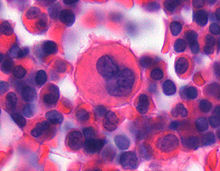
Megakaryocytes are the largest cells in the bone marrow, their diameter is 35–150 µm. The nucleus is irregularly lobed, contains coarse-grained chromatin and no visible nucleoli . Under the light microscope, it can appear as if there are several cell nuclei.
The cytoplasm contains numerous mitochondria , a well-developed endoplasmic reticulum , an extensive Golgi apparatus, and a large number of ribosomes . It can be found also granules , as well as those found in platelets. They are divided into α-granules, electron-dense granules and lysosomes and contain the proteins and substances that are important for platelet function, including ADP , ATP , calcium , growth and clotting factors .
Occurrence
The total number of megakaryocytes is about 6 million per kg of body weight. The majority of megakaryocytes are found in the red bone marrow . At around 1%, however, they only make up a small proportion of the cells living there.
In small numbers (five to seven per milliliter) they are also found in the circulating blood, but are then largely, but not completely, filtered out in the pulmonary capillaries. However, these circulating cells then contain almost no cytoplasm. The number of circulating megakaryocytes is increased in newborns and young children as well as in mothers immediately after birth, as well as in infections, inflammations, malignant neoplasms, disseminated intravascular coagulation and myeloproliferative diseases .
Megakaryopoiesis
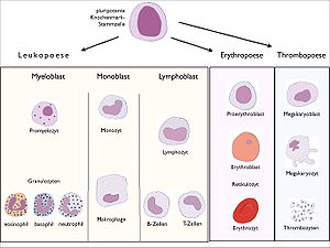
As megakaryopoiesis the development of megakaryocytes from specific is stem cells ( Hämozytoblasten ) referred to in the bone marrow. It lasts about 10 days.
Under the influence of thrombopoietin (TPO), the pluripotent stem cells in the bone marrow form megakaryoblasts through mitotic division . These have a diameter of 20 to 30 µm, a basophilic cytoplasm and numerous nucleoli in the cell nucleus. They no longer have the ability to divide, but can still multiply their DNA ( endomitosis ). After several of these endomitoses, they are polyploid and ultimately contain up to 64 complete sets of chromosomes (64N). By definition, megakaryoblasts do not contain any granulation in the cytoplasm. This only occurs at the next stage of maturation, the promegakaryocytes . As the development progresses, the basophilia of the cytoplasm decreases while the azure granulation increases. The volume of the cytoplasm increases relative to the volume of the cell nucleus. DNA synthesis comes to a standstill. The mature megakaryocyte is usually very cytoplasmic with azurophilic cytoplasm.
Thrombopoiesis
Thrombopoiesis refers to the formation of thrombocytes (blood platelets) by megakaryocytes. One megakaryocyte can produce 1,000 to 3,000 platelets.
After the cytoplasm of the megakaryocyte has matured , it changes into so-called proplatelets . Several thick extensions, the pseudopodia, are formed that penetrate the sinusoids of the bone marrow. There they form cord-like constrictions in which platelet material is found that was transported there along microfilaments from the center of the cell. Completely constricted, they form the blood platelets. So far, however, it is not known whether the platelets are fully developed and released into the bloodstream or whether the formation is only completed in the vessels.
Diseases
In essential thrombocythemia , the megakaryocytes release an uncontrolled high number of thrombocytes into the bloodstream, so that they reach a concentration of up to 500,000 per µl (normal value: 150,000–350,000 / µl). The concentration of the other blood components, however, is normal. Essential thrombocythemia is caused by an increased sensitivity of the megakaryocytes to thrombopoietin. As a result, there are increased numbers of abnormally large, mature megakaryocytes in the bone marrow. When making the diagnosis, however, other causes of thrombocytosis , such as infections, anemia, or other diseases of the bone marrow, must be ruled out.
In thrombocytopenia , the number of circulating platelets is reduced. In addition to a multitude of possible causes outside the bone marrow, the disease can also be caused by impaired formation in the bone marrow. In an aplastic disorder of the bone marrow, the number of megakaryocytes is reduced or completely absent. This is either due to a genetic disorder such as Fanconi anemia or it can be acquired. These include damage to the bone marrow through poisoning or radiation, bone marrow infiltration through malignant tumors, displacement through uncontrolled proliferation of cells in leukemia or a sclerotherapy of the blood-forming bone marrow osteomyelofibrosis . Furthermore, maturation disorders due to a lack of vitamin B 12 or folic acid can reversibly reduce the number of mature, functional megakaryocytes.
swell
literature
- Tinsley R. Harrison et al .: Harrison's Principles of Internal Medicine . Mcgraw-Hill Professional, 2005, ISBN 0-07-007272-8 .
- G. Herold and others: Internal medicine. 2006.
Individual evidence
- ↑ Barbara J. Bain: Blood Cells. A practical guide . Blackwell Publishing, 2006, ISBN 1-4051-4265-0 .
- ↑ Peter C. Heinrich, Matthias Mueller, Lutz Graeve (eds.): Löffler / Petrides Biochemistry and Pathobiochemistry . 9th edition. Springer-Verlag Berlin Heidelberg, 2014, ISBN 978-3-642-17971-6 , p. 878 .
- ↑ O. Benke, A. Forer: From megakaryocytes to platelets: platelet morphogenesis takes place in the bloodstream. In: Eur J Haematol Suppl. 61, 1998, pp. 3-23. PMID 9658684

