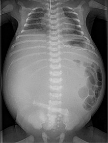
Meconium peritonitis
| Classification according to ICD-10 | |
|---|---|
| P78.0 | Intestinal perforation in the perinatal period - meconium peritonitis |
| ICD-10 online (WHO version 2019) | |

A meconium peritonitis arises when there is in a child before birth to a bowel perforation occurs and so meconium , so the intestinal contents of the unborn child, in the abdominal cavity is excreted. It usually develops during the second half of the fetal period, but can rarely occur after birth.
frequency
The frequency is given as 1 in 35,000 live births.
causes
In 10–15% of cases the underlying condition is cystic fibrosis , in which the abnormal meconium displaces the small intestine (at the level of the ileum ) and expands the intestinal wall to the point of perforation.
Other causes are:
- primary small intestinal atresia
- severe bowel stenosis
- Necrosis due to ischemia of the intestinal wall
- Perforation of a Meckel's diverticulum
- Volvulus
- Intussusception
- Adhesions
- internal hernias
- Hirschsprung's disease
- Colon atresia
clinic
Since the intestine has not yet been colonized by bacteria, the resulting inflammation of the abdominal cavity is sterile as a foreign body reaction. Due to the rapid onset of calcium deposits, characteristic delicate linear calcifications can be found in the x-ray of the newborn.
A pseudocyst can also develop, which depicts a mass that more or less displaces the intestine, depending on its size. If the perforation persists until after the birth, gas can also escape and a bacterial inflammation can develop.
classification
Three types can be distinguished:
- Generalized peritonitis
- Cyst formation
- Formation of adhesions (adhesions)
Diagnosis
Using ultrasound presumption diagnosis can be made in many cases already in the womb. Significant findings are polyhydramnios , intestinal loop dilation, ascites or pseudocysts .
therapy and progress
The perforation site can close spontaneously, so that surgery is not necessary in all cases. The extent of the existing inflammatory reaction has a decisive influence on the course.
history
The first description of the disease is said to have been made by Giovanni Battista Morgagni in 1761 .
The typical radiological findings were published in 1944 by E. Neuhauser .
Individual evidence
- ↑ a b M. Bettex, N. Genton, M. Stockmann (Ed.): Pediatric Surgery. Diagnostics, indication, therapy, prognosis. 2nd edition, Thieme 1982, p. 7.61, ISBN 978-3-642-11330-7
- ^ WS Lorimer, DG Ellis: Meconium peritonitis. In: Surgery. Vol. 60, No. 2, August 1966, pp. 470-475, PMID 5920370 .
- ↑ K. Uchida, Y. Koike, K. Matsushita, Y. Nagano, K. Hashimoto, K. Otake, M. Inoue, M. Kusunoki: Meconium peritonitis: Prenatal diagnosis of a rare entity and postnatal management. In: Intractable & rare diseases research. Vol. 4, No. 2, May 2015, pp. 93-97, doi: 10.5582 / irdr.2015.01011 , PMID 25984428 , PMC 4428193 (free full text).
- ↑ D. Möslinger, K. Chalubinski, M. Radner, M. Weninger, A. Rokitansky, G. Bernaschek, A. Pollak: [Meconium peritonitis: intrauterine follow-up-postnatal outcome]. In: Wiener Klinische Wochenschrift. Vol. 107, No. 4, 1995, pp. 141-145, PMID 7709630 .
- ^ Giovanni Battista Morgagni: De sedibus et causis morborum per anatomists indagatis . 1761 ( archive.org ).
- ↑ EBD Neuhauser: The roentgendiagnosis of fetal moeconium peritonitis. In: American Journal of Roentgenology Vol. 51, 1944, p. 421
Sources and web links
- A. Zink and P. Sacher: Pre- and postnatal imaging diagnostics of meconium peritonitis; Surgical Clinic, University Children's Hospital Zurich Switzerland on http://www.kinderchirurgie.ch/
- W. Schuster, D. Färber (Ed.): Children's radiology. Imaging diagnostics. Springer 1996, Vol. 2, p. 546, ISBN 3-540-60224-0 .