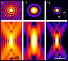
Point spread function

The point spread , point spread function , point spread function , point spread function or point spread function (engl. "Point spread function", shortly PSF ) in describing high-frequency technology , optics and image processing , the effect of band-limiting factors such as:
- Diffraction phenomena on apertures
- Image errors
- Influence of the sensor area or aperture
It indicates how an idealized, point-like object would be mapped by a system. Often the shape of the point response is independent of the original location of the ideal, point-like object. In this case one speaks of a linear system and the overall response of the system can be calculated as the sum of the point responses of the object broken down into its points.
In light microscopy , the maximum achievable resolution of an objective , which is limited by diffraction , can be determined with the aid of the PSF. The resolution of a microscope is generally understood to be the distance that two point-like structures must have so that they can be recognized as separate structures (and not as one structure).
However, usually only the image of a small spherical structure is recorded microscopically, for example the microscopic image of very small fluorescent latex spheres. In order to determine a measure of the achievable resolution in the x, y or z direction, the brightness distribution along a profile through the brightest point parallel to the desired axis is first determined. The width of this brightness distribution halfway between the intensity maximum and intensity minimum (FWHM Full Width Half Maximum ) is often given as the achievable resolution. Alternatively, the point spread function can also be calculated theoretically for various experimental mapping scenarios.
While the width (FWHM) of the point spread function is the most important factor for resolution, the resolution actually achieved by a microscopic image also depends on other factors such as the signal-to-noise ratio .
See also
- Convolution (mathematics) Building an image by summing the point responses of the decomposed object corresponds mathematically to a convolution
- Impulse response
- Modulation transfer function
Web links
- PSF Generator Free program for calculating point spread functions at the École polytechnique fédérale de Lausanne (in English)
- Non-stationary Correction of Optical Aberrations Paper, in which PSF are used to correct aberrations in lenses.
Individual evidence
- ^ Nasse MJ, Woehl JC, Huant S .: High-resolution mapping of the three-dimensional point spread function in the near-focus region of a confocal microscope . In: Appl. Phys. Lett. . 90, No. 031106, 2007, pp. 031106-1-3. doi : 10.1063 / 1.2431764 .
-
^ B. Richards, E. Wolf: Electromagnetic diffraction in optical systems II. Structure of the image field in an aplanatic system . In: Proc. R. Soc. Lond. A . Vol. 253, 1959, pp. 358-379 , JSTOR : 100740 . Nasse MJ, Woehl JC: Realistic modeling of the illumination point spread function in confocal scanning optical microscopy . In: J. Opt. Soc. At the. A . 27, No. 2, 2010, pp. 295-302. doi : 10.1364 / JOSAA.27.000295 .