Human flea
| Human flea | ||||||||||||
|---|---|---|---|---|---|---|---|---|---|---|---|---|

The human flea ( Pulex irritans ), illustration from Medical and Veterinary Entomology (1915) |
||||||||||||
| Systematics | ||||||||||||
|
||||||||||||
| Scientific name | ||||||||||||
| Pulex irritans | ||||||||||||
| Linnaeus , 1758 |
The human flea ( Pulex irritans ), often just shortened to the flea , is a blood-sucking insect belonging to the order of the fleas (Siphonaptera).
features


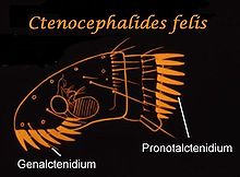
The human flea has the general body shape of the fleas and is difficult to distinguish from other flea species at first glance. Like most fleas, it is yellowish to yellowish brown in color, wingless with jump bones, laterally flattened with a very stable exoskeleton (heavily sclerotized ). Females of the species reach a body length of about 2.5 to 3.5 millimeters, the slightly smaller males only 2 to 2.5 millimeters. Unlike many other fleas lacking both on the cheeks ( Genae ), the lower edge of the head below the eyes, as well as at the back edge of the throat plate ( pronotum ) tooth combs or ctenidia that are eye-catching series of very strong, mostly triangular shaped mandrel bristles . The shape of the antenna, which is difficult to see, is important for the exact definition of the species . These are short and can be placed in a pit behind the eye. In the genus Pulex , the end lobe of the antennae is asymmetrically shaped, its first limb widened in the shape of a leaf, its tip protruding beyond the edge of the eye.
The human flea can be differentiated from other flea species without ctenidia, which occur widespread in connection with humans and their domestic animals: In the chicken flea (or also chicken comb flea) Echidnophaga gallinacea the front edge of the head is angular when viewed from the side, not rounded like in the human flea. It is more difficult to distinguish it from the rat flea or plague flea ( Xenopsylla cheopis ), which, however, occurs only rarely in Central Europe. In this case, the mesopleuras (a sclerite on the side of the middle trunk segment) are apparently divided into two by a reinforced ridge, and the eyepiece bristle, a small bristle in the area of the eyes, is located in front of the conspicuous, button-shaped eye, not below it as in the human flea . The genus Tunga , with the sand flea Tunga penetrans , is similar. In the case of the human flea, the trunk segments are roughly the same size when viewed from the side, and the antenna of Tunga is shorter, its tip does not protrude beyond the eye.
Occurrence
The human flea occurs worldwide. Since all related species of the genus Pulex live in America and no ancient fossil or subfossil remains have been found from most of the world, the prevailing scientific view is that the species was originally only distributed in America and was only introduced worldwide by humans. A widely accepted model sees the transition to humans in South America, where humans came into closer contact with the parasite for the first time through the domestication of guinea pigs ( Cavia porcellus ). From here, through cultural contact with groups of people, it would have been brought into the Old World via the Bering Strait (or possibly elsewhere via boat traffic) and spread there. The human flea is one of the mobile flea species which, although they remain dependent on a bed, burrow or nest for the larvae to reproduce and develop, live as adults as ectoparasites on their host and can therefore be easily carried away by the host as soon as there is direct contact .
Flea remains are rather rare in archaeological excavations. They are from pre-Columbian Indian graves from southern Peru, which date back to around 1000 BC. To be dated. If one takes the domestication of the guinea pig, which possibly already took place 7000 BC, but probably no later than 5000 BC, as a starting point, it is remarkable that the first evidence of human flea from the European Neolithic , around 3000 BC Old world present.
Also noteworthy is the find in the workers' settlement in Amarna , Egypt , which was only inhabited for about 25 years, between about 1350 and 1323 BC. The remains of 35 human fleas (and one cat flea) were found in a burial ground, which proves the widespread presence of the species in North Africa as early as the end of the 2nd millennium BC.
Since humans are the only species of primate that are infested by fleas, it is extremely unlikely that humans can be the parasite's primary host, so the term “humans” fleas is misleading. As hosts next to humans and rodents, guinea pigs are known: pigs, for example also the South American umbilical pigs (peccaries) and domestic pigs, numerous predators, especially foxes ; in the red fox ( Vulpes vulpes ) it is the second most common flea, and in the American swift fox ( Vulpes velox ) the most common. Overall, a large number of at least occasionally infected host species are known, including numerous rodents, notably the (cave-nesting) owl ( Athene cunicularia ), a bird species.
The human flea is also widespread on domestic animals; in addition to domestic pigs, domestic goats and, rarely, domestic cats have also been identified as hosts. Alongside pigs, domestic dogs are one of the most important hosts: around 10 percent of dogs worldwide are said to be infected with this species.
The human flea has become rare in Central Europe. Much more often people are attacked by cat fleas ( Ctenocephalides felis ) or dog fleas ( Ctenocephalides canis ).
nutrition
It sucks blood for food, but it can go without a meal for up to a year. Warm, humid regions on the body are preferred for the sting. A single flea can usually cover the whole body with stings in a short time at night. Usually, the flea will eat one blood meal a day. If possible, he often takes on twenty times his own weight. Some of the thickened blood is excreted shortly afterwards.
The flea bites are occasionally arranged in a row; one also speaks of flea street .
development
The development of the human flea proceeds through the stages egg, larva, pupa and imago . Such a cycle usually lasts from a few weeks to eight months.
Egg laying
The first mating takes place about 8 to 24 hours after eating. The female fleas begin to lay eggs about one day after mating. A female lays around 50 eggs per day, which are laid indiscriminately on the host organism. They are soft, oval, light, only about ½ mm in size and have no sticky outer shell, which is why they can fall off the host body at any time.
Young larvae
The young larvae hatch about 2 to 14 days after egg-laying and prefer to hide in carpets, on floors, especially in the corners and wall areas near the heater, in upholstered furniture, pillows, mats and mattresses. The blood that has been thickened and excreted by a flea serves as food for the 5 mm long, white, thread-thin larvae, as they cannot yet suckle.
Harmful effect
As a typical reaction to a flea bite, small papules develop in humans . These are red in color, are usually hard, slightly raised and more or less itchy. Scratching these papules can cause secondary infections.
Human fleas as carriers of disease
The human flea can occasionally mechanically transmit the causative agents of typhus and bubonic plague when sucking blood . The transmission occurs through the contact of the fleas or the contaminated flea body ( proboscis ) with the stab wound. Furthermore, human fleas can be intermediate hosts for various tapeworm species such as B. be the cucumber seed tapeworm ( Dipylidium caninum ) and transmit them.
A group of researchers at the University of Marseille led by Didier Raoult is of the opinion that when the host changes, the human flea, as well as the head louse ( Pediculus humanus capitis ) and the body louse ( Pediculus humanus humanus ), can also be considered as vectors of the plague , as all of these parasites Can absorb plague bacteria. Some researchers question the entire model, which was at times widely accepted as the standard, that the plague was transmitted to humans by the house rat and its parasite rat flea ( Xenopsylla cheopis ), and these constituted the essential reservoir for the survival of the plague pathogen between epidemics . According to their conclusions, the human flea would have been the main vector of the plague in ancient and medieval Europe, while the rat flea was more important in Central and East Asia. One difficulty with the hypothesis is that the pathogen from human flea is much less easily transferred to humans than from rat flea, because the intestine does not become blocked, which means that large amounts of bacteria pass into the blood during the stinging process. However, its importance as a vector for the disease was made very likely for regions where the plague is still endemic today, such as African Tanzania. It is significant that human fleas can transmit the disease directly from person to person; an intermediate host such as the house rat is not required.
Web links
Individual evidence
- ↑ Hubert Schumann: Fleas (Siphonaptera) . In: Hans-Joachim Hannemann, Bernhard Klausnitzer, Konrad Senglaub (eds.): Stresemann excursion fauna . 7th edition. tape 2/2 : invertebrates . People and knowledge, Berlin 1990, ISBN 3-06-012558-9 , pp. 300 .
- ^ Frans GAM Smit: Siphonaptera . In: Royal Entomological Society of London (Ed.): Handbook for the identification of British insects . tape 1 , no. 16 . Self-published, London 1957, p. 1-91 .
- ↑ Gunvor Brink-Lindroth, Frans GAM Smit: The fleas (Siphonaptera) of Fennoscandia and Denmark . In: Fauna Entomologica Scandinavica . tape 41 . Brill, Leiden / Boston 2007, ISBN 978-90-474-2075-0 , pp. 137 ff .
- ↑ Harry D. Pratt, John S. Wiseman: Fleas of public health importance . In: US Department of Health, Education, and Welfare (Ed.): Public health series . No. 772 . US Government Printing Office, Washington 1962, p. 17 .
- ^ E. Fred Legner: Key to Siphonaptera of Medical Importance. University of California.
- ^ Richard C. Russell, Domenico Otranto, Richard L. Wall: The Encyclopedia of Medical and Veterinary Entomology. CABI, Wallingford OX / Boston MA 2013, ISBN 978-1-78064-037-2 , p. 117.
- ^ Paul C. Buckland, Jon P. Sadler: A Biogeography of the Human Flea, Pulex irritans L. (Siphonaptera: Pulicidae) . In: Journal of Biogeography . tape 16 , no. 2 , 1989, pp. 115-120 , JSTOR : 2845085 .
- ^ Eva Panagiotakopulu, Paul C. Buckland: A thousand bites - Insect introductions and late Holocene environments . In: Quaternary Science Reviews . tape 156 , 2017, p. 25-35 , doi : 10.1016 / j.quascirev.2016.11.014 .
- ^ Eva Panagiotakopulu: Fleas from pharaonic Amarna . In: Antiquity . tape 75 , 2001, p. 499-500 .
- ^ Eva Panagiotakopulu: Pharaonic Egypt and the origins of plague . In: Journal of Biogeography . tape 31 , no. 2 , 2004, p. 269-275 , doi : 10.1046 / j.0305-0270.2003.01009.x .
- ^ A b Gerhard Dobler, Martin Pfeffer: Fleas as parasites of the family Canidae . In: Parasites & Vectors . tape 4 , no. 139 , 2011, pp. 1-11 , doi : 10.1186 / 1756-3305-4-139 .
- ↑ Hans-Jürgen von der Burchard: Mosquitoes - The Springer Elite . On: planet-wissen.de as of June 1, 2009, accessed on July 4, 2012.
- ↑ D. Raoult et al .: Experimental model to evaluate the human body louse as a vector of plague. In: The Journal of Infectious Diseases . Volume 194, No. 11, Dec. 2006, pp. 1589-1596, PMID 17083045 .
- ^ D. Raoult et al .: Body lice, yersinia pestis orientalis, and black death. In: Emerging Infectious Diseases . Volume 16, No. 5, May 2010, pp. 892-893, PMID 20409400 .
- ↑ Anne Karin Hufthammer, Lars Walløe: Rats can not havebeen intermediate hosts for Yersinia pestis during medieval plague epidemics in Northern Europe . In: Journal of Archaeological Science . tape 40 , 2013, p. 1752–1759 , doi : 10.1016 / j.jas.2012.12.007 .
- Jump up ↑ Anne Laudisoit, Herwig Leirs, Rhodes H. Makundi, Stefan Van Dongen, Stephen Davis, Simon Neerinckx, Jozef Deckers, Roland Libois: Plague and the Human Flea, Tanzania . In: Emerging Infectious Diseases . tape 13 , no. 5 , 2007, p. 687-693 ., Doi : 10.3201 / eid1305.061084 .