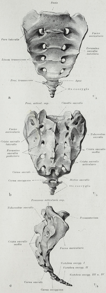
Sacrum
The sacrum ( lat. Os sacrum , sacrum ) is a bone of the terrestrial vertebrates . In the adult human, it is roughly wedge-shaped. It develops through fusion ( synostosis ) from the originally individual, converging vertebrae , the cross vertebrae (sacral vertebrae). In adolescents, this fusion (previously a cartilaginous bond) takes place until the end of the growth phase. The sacrum encloses the rear portion of the spinal canal and forms with two hip bones of the pelvic girdle .
Vortex number
The number of fused vertebrae varies within the mammalian class between three (e.g. dogs ) and five ( humans , horses ). The individual vertebrae can still be recognized by fused lines ( Lineae transversae ).
In a few people, the upper original sacral vertebra (S1) is not fused with the other sacral vertebrae as usual ( lumbarization ). So these people seem to have six lumbar vertebrae instead of five. In any case, this results in greater mobility of the spine and can mean a lower load-bearing capacity of the spine, but usually does not lead to complaints. In other cases, the 5th lumbar vertebra is wholly or partially fused with the sacrum ( sacralization ). This can cause scoliosis and neurological damage from narrowing of the intervertebral hole.
Caudally (towards the tail) - i.e. backwards in animals and downwards in humans - the tailbone ( Os coccygis ) or the caudal spine connects .
Exiting nerves
The spinal nerves emerging from the sacrum form a network together with parts of the nerves emerging from the lower lumbar vertebrae - the lumbosacral plexus . From this network, nerves are formed that primarily supply the pelvis and legs.
anatomy
Despite the fusion, the sacrum still has all of the basic characteristics of the vertebrae.
The former spinous processes form a crest that differs in height, the crista sacralis mediana . Only in horses each, very long spinous processes (usually remain spinous processes ) isolated.
Upwards (in animals in front) lies on a small extension ( processus articularis cranialis ) on both sides a joint surface to the last lumbar vertebra. The remaining articular processes form in some species (e.g. humans, ruminants) a ridge-like elevation ( Crista sacralis intermedia ).
In all mammals, the transverse processes form a broad plate, the pars lateralis ("lateral part"). The anterior part of the lateral part is enlarged to form the sacral wing ( ala ossis sacri ) in animals . In all mammals (including humans) there is an ear-shaped joint surface ( facies auricularis ) on both sides , which together with the joint surface of the same name on the iliac bone form the respective sacroiliac joint (sacrum-iliac joint). They attach the pelvis to the torso . The lateral edge of the pars lateralis is called the lateral sacral crest .
The pars lateralis divides the openings for the spinal cord nerves . The foramina sacralia posteriora (animals: Foramina sacralia dorsalia ), which are used by the dorsal branches of the sacral spinal cord nerves to exit, and the foramina sacralia anteriora (animals: Foramina sacralia pelvina ) for the corresponding ventral branches can be found on the back of each .
particularities
The sacrum arcuatum is a particularly strongly curved sacrum.
Movement possibilities
The movement of the sacrum is called nutation or counter- nutation . Here, the displaced promontory to ventrocaudal (downward and forward) and the lower surface ( Apex sacrum ), the to the tailbone ( coccyx ) followed, after dorsocranially (to the rear and above). The sacrum-iliac joint ( sacroiliac joint ) not only rotates, but also shifts the surfaces against each other.
In cranio-sacral therapy , several axes of movement are postulated, the upper transverse axis (OTA), the middle transverse axis (MTA) - nutation and counter-nutation -, the lower transverse axis (UTA), a cranio-caudal axis and a right and left diagonal axis .
Web links
Individual evidence
- ↑ a b c d e Franz-Viktor Salomon: Kreuzwirbel, Vetrebrae sacrales . In: Franz-Viktor Salomon et al. (Ed.): Anatomy for veterinary medicine . 2nd Edition. Enke, Stuttgart 2008, ISBN 3-8304-1007-7 , pp. 45-46 .
- ↑ Jochen Fanghänel et al .: Waldeyer - Anatomie des Menschen . Walter de Gruyter, 18th edition 2009, ISBN 9783110911190 , pp. 637–638.
- ^ A b c Johannes W. Rohen, Elke Lütjen-Drecoll: Functional anatomy of humans: textbook of macroscopic anatomy according to functional aspects. Schattauer Verlag, 2006, ISBN 9783794524402 , p. 42.


