
White matter
As a white substance (WS), or latin substantia alba to portions of the designated central nervous system , which consists predominantly of pathways or nerve fibers exist and thus primarily axons included. These are contrasted with gray matter as those parts that mainly contain nerve cell bodies ( perikarya ) and consist, for example, of nuclei or core areas. The macroscopically visible white color is caused by enveloping glial cells or the myelin sheaths of the nerve fibers.
The white matter lies on the outside of the spinal cord , surrounding its gray matter, and is macroscopically divided into anterior ( anterior funiculus ), lateral ( lateral funiculus ) and posterior ( funiculus posterior ) cord .
In the cerebral cortex , on the other hand, the gray matter is on the outside and surrounds the white matter of the medullary bed . In the reticular formation of the brainstem, on the other hand, it is difficult to distinguish. Localized clusters of nerve cell bodies are used as nuclei ( Nuclei referred to); they can also be embedded in areas of the white matter or vice versa, such as in the solitary tract .
White Matter Lesion
When white matter lesion (WML) refers to damage to the white matter, which occur quite frequently with increasing age in the general population and those with cognitive impairment and stroke are connected. They are also known as white matter hyperintensities because they appear as bright white spots in MRI images. In the Rotterdam Scan Study of 1077 non-demented people between the ages of 60 and 89 years, only 5% did not find WML. 80% had periventricular WML, which is associated with cognitive and psychomotor disorders.
Histopathologically , WML is described as gliosis, demyelination, and loss of axons and diagnosed as leucoaraiosis or vascular dementia .
See also
gallery

Cross section of the spinal cord: white matter outside and gray matter inside.
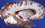
Sagittal section of the brain: gray matter outside and white matter inside.

