pathology


The pathology (from ancient Greek πάθος páthos , German , sickness, suffering ' and λόγος , lógos , German , teaching' , ie "doctrine of suffering") is the study of the abnormal and pathological processes and states in the body and their causes. The research covers both individual phenomena ( symptoms ) and groups of symptoms ( syndromes ) as well as malformations of all kinds. The pathology examines the origin ( etiology ), the mode of development ( pathogenesis ), the course and the effects of diseases including the respective processes in the body ( functional Pathology or pathophysiology ).
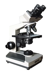
Pathological diagnostics , i.e. the work of the pathologist (specialist in pathology), is primarily based on the assessment of tissues on the basis of their macroscopic (pathological anatomy ) and light microscopic aspects ( histopathology , cytology ). Biochemical and molecular biological methods are increasingly being used, electron microscopy in research . Pathologists also perform clinical autopsies . However, the examination of the tissues of living patients ( biopsy ) predominates by far. The term clinical pathology is occasionally used for this "pathology in the living" .
To the subject
The Greek term παθολογία pathologia can be derived from the words πάθος páthos 'disease, suffering, passion' and λόγος lógos 'word, sense, reason, teaching'. The Latinized noun pathologia as 'the doctrine of suffering' or 'disease theory' has only been documented since the 16th century, originated from the Greek pathologikè téchne ('knowledge of disease') and goes back to the expression pathologikós in Galenos , who means "Person who is knowledgeable in the scientific handling of illness" had designated. The adjective pathological means "pathological". In the first half of the 19th century, general pathology or French pathology générale was the "general natural science of diseases", something like an explanation of disease plus nosology .
Concepts such as psychopathology , pathological play , pathological science and pathological example have nothing to do with anatomy .
history
In its current form, pathology, already popularized as a word by Jean Fernel , goes back to the Italian researcher Giovanni Battista Morgagni (1682–1771), who wrote his five-volume work De sedibus et causis morborum (“From the seat and the causes of diseases “) Laid the foundation stone for scientific research in 1761 and is particularly considered to be the founder of pathological anatomy.
In ancient times, cadavers were opened in Egypt and Greece , but they were more used for anatomical education. It was not until the end of the 18th century that, due to the increasing understanding of the importance of the post- mortem examination, the first specialist representatives who were specifically responsible for the sections were appointed. The first so-called prosector (lat. Prosecare : pre-cutting ) began its work in 1796 at the Vienna General Hospital. The first chair for pathology was established in Strasbourg in 1819 ( Jean-Frédéric Lobstein , 1777-1835). Pathology was introduced as an examination subject in Vienna in 1844.
Analytical pathology, which opposed the theoretical concepts of his time and was based on empirical methods, was founded around 1840 by the Italian doctor Maurizio Bufalini (1787–1885).
Pathological anatomy , one of the pioneers of the Italian anatomist and pathologist Antonio Benivieni at the end of the 15th century, established itself as an independent subject at German universities between 1845 and 1876. The first American work in this field was published by the anatomist William Edmonds Horner in 1829. From 1858 onwards, Rudolf Virchow , for whom the first pathological institute in Germany he directed was established in Berlin in 1856, made known the cellular pathology he had developed in Würzburg , which now examined pathological changes on the level of body cells. This is a main component of today's disease concept . Virchow is considered to be the "initiator of modern pathology in German-speaking countries". Due to the influence of Virchow's works, the term pathology replaced the sub-area designation "pathological anatomy" in the German-speaking world . Historical research into the development and fundamentals of pathology also began in the 19th century.
General and Special Pathology
For academic teaching ("Pathology explains diseases") a distinction is made between general pathology and special pathology:
- The General Pathology is a general pathology that on the etiology (medicine) , the causal (why?) And formal (how?) Pathogenesis as well as the concept of disease (What is disease?) Provides information.
- Adaptation reactions (adaptation)
- necrosis
- General circulatory disorders
- Thrombosis , embolism , infarction
- Inflammation , acute and chronic
- Immunopathology (allergy, transplantation)
- Tumors
- The special pathology describes the special diseases of the organs based on macroscopic, microscopic, electron microscopic, immunohistological and molecular pathological findings. When viewed together with the associated clinical findings, the clinicopathological diagnosis required for the further treatment of a patient is obtained for the individual case of illness.
- Atherosclerosis
- Myocardial infarction
- Other heart diseases
- Respiratory diseases
- Digestive tract and liver
- Biliary tract and pancreas
- Kidney disease
- Lower urinary tract
- Male and female reproductive organs
- Female Breast Disorders
- Skeletal diseases , bone tumors
- Central nervous system disorders
Duties of the pathologist
Examination of tissue and cell samples
After surgical removal of an organ or removal of a small piece of tissue or cell samples ( cytodiagnostics ) by a doctor, the corresponding tissue is examined by the pathologist. Small biopsies are processed directly into slices that can be viewed under the microscope . Large specimens are first prepared and assessed with the naked eye (macroscopically). Conspicuous components with possible pathological changes are cut out of the specimen (so-called "cut") and then processed by the laboratory to make cut specimens. The quick cut is a special form of cutting . Here, frozen sections of tissue are made intraoperatively (during an operation in which the patient is still under anesthesia) , e.g. B. a resection margin in a tumor operation. Since frozen sections are generally of poor quality and often do not allow further examinations, paraffin sections with HE staining are made as standard outside of quick section situations .
With the help of the microscope, the pathologist provides information about the type of disease and its severity. He thus makes diagnoses that cannot be made by a clinical or radiological examination alone . In the case of a tumor and the question of whether it is benign or malignant , a pathologist is needed. He examines the type, the size, the extent, the malignancy of a tumor and checks whether it was removed in the normal during the operation. It thus provides the clinical doctor with many important prognostic factors (e.g. TNM classification ) that are indispensable for the correct treatment of the patient. In addition to histological assessment, highly specialized methods such as immunohistochemistry or molecular pathology (e.g. fluorescence in situ hybridization , PCR ) are also used in modern pathology . This allows information about a tumor to be obtained at the molecular level, which is crucial for a certain form of therapy (e.g. hormone receptors in breast cancer as the basis for treatment with tamoxifen ).
autopsy
Another task of the pathologist is to carry out autopsies, which is why pathology is often confused with forensic medicine . An autopsy is carried out by the pathologist if a patient has died of natural causes (e.g. after a heart attack) and his relatives agree to the autopsy. This so-called clinical autopsy serves to clarify the cause of death and the pre-existing diseases. It gives the attending physician feedback on the correctness of his diagnosis and treatment. Such clarification of the cause of death can often be a relief for the relatives and free them from self-reproach (e.g. after the sudden onset of a fatal course of the disease). An autopsy can also provide information about familial risk factors (e.g. types of cancer or hereditary diseases). Forensic medicine, on the other hand, deals among other things with the clarification of unnatural causes of death (e.g. murder or accident ). Both pathologists and forensic doctors are annoyed when TV crime novels and in common parlance always only refer to “pathologists” or “pathology”, but when it is generally about a forensic doctor. The common mistake is explained by a mistranslation: in American usage, the forensic pathologist corresponds to the forensic pathologist .
Although most lay people think of pathology as autopsies, the work of the pathologist nowadays primarily serves the living patient. With his histological examinations he makes an important contribution to the correct treatment. In modern pathology, few autopsies (depending on the institute between 0 and 200 per year) stand in the way of tens of thousands of biopsies from living patients.
quality control
Pathology continues to be one of the most important instruments of quality assurance in medicine . In order to maintain and improve the medical standard, a collegial confrontation between the clinician and the diagnostic diagnostics of the pathologist is often required, not only during the life of the patient, but also after his death . The corresponding event has a permanent place as a "clinical pathological conference" not only in everyday clinical practice, where weekly meetings often take place where pathologists discuss their findings with the doctors working at the patient's bedside. As a "team player", the pathologist is particularly involved in interdisciplinary tumor conferences, where, together with radiologists, oncologists and other disciplines, the course is set for the individually tailored therapy of the patient.
Teaching and Research
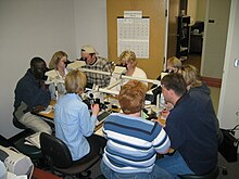
At university hospitals in particular , pathologists are also involved in the training of young doctors and in research.
Sub-areas of diagnostic pathology
The activity of the pathologist is divided into different work areas.
- Pathological anatomy (macroscopy): The examination of pathological changes in the body with the naked eye, for example as part of an autopsy or cutting, i.e. H. in the preparation of surgical specimens. As with the physical examination of the living, many conclusions can be drawn about the disease process.
-
Histopathology : The examination of tissue with the light microscope is the essential activity of the pathologist and often the " gold standard " for diagnosis, especially of tumor diseases. For this purpose, after fixation in formalin and embedding in paraffin, thin sections are prepared, colored and usually assessed at 10 to 400 times magnification of the tissue to be examined. HE staining has established itself as the standard staining , which is often sufficient for diagnosis. In addition, there are many special stains ( histochemistry ) that emphasize special tissue properties, e.g. B .:
- Masson-Goldner - connective tissue green
- Elastica-van-Gieson (EvG) - connective tissue red and elastic fibers purple
- Congo red - amyloid red
- PAS - carbohydrates / glycoproteins red, including glycogen , mucins, mushrooms, plant material, basement membrane
- Polarization : Using a polarization filter, microscopic substances can be made to glow, e.g. B. collagen, amyloid (after Congo red staining), foreign material or uric acid crystals (native preparation).
- Rapid section diagnostics : The tissue is frozen, sectioned and HE-stained directly in order to e.g. B. to make a quick preliminary statement during an operation. The disadvantage is the higher workload per case and the lower accuracy due to freezing artifacts and pure HE diagnostics.
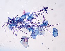
- Cytopathology : Examination of single cells ( cytodiagnostics ) instead of tissue samples. The best known cytological procedure is the so-called PAP smear from the cervix for the early detection of cervical cancer . Furthermore, body fluids such as urine, effusions or bronchial secretions can be examined. Cytological examinations are quick, inexpensive and less invasive to perform than biopsies, but they are also not always particularly meaningful. They are suitable as an addiction test ( screening ), but cannot usually replace a biopsy for a definitive diagnosis.
- Immunohistochemistry and immunocytology : Specific protein structures (such as DNA repair proteins, cytoskeletal proteins or receptors) of the cells to be examined can be made visiblewith dye-labeled antibodies. This technology has revolutionized tumor diagnostics in particular. Tumors can thus z. B. can be typed more precisely with regard to their type diagnosis (e.g. lymphoma ), metastases can be better assigned to their origin and prognosis and therapy-relevant markers can be determined, such as B. in breast cancer the growth rate ( Mib1 / Ki67 ), the estrogen receptor , the progesterone receptor and HER2 / neu .
- The electron microscopy plays a subordinate role. It is used for certain questions, such as B. ciliary diseases ( Kartagener syndrome ) or certain kidney diseases ( glomerulonephritis ).
- Molecular pathology : The youngest branch of pathology examines changes at the genetic or DNA or RNA level e.g. B. by means of in-situ hybridization (ISH), PCR , sequencing , etc. B. tumor-specific mutations can be detected such. B. SYT-SSX translocation in synovial sarcoma, MDM2 and CDK4 amplification in liposarcoma, and ESW-FLI1 translocation in Ewing sarcoma . Or therapy-relevant mutations can be examined, such as B. in the BRAF gene in malignant melanoma or in the EGFR gene in lung cancer .
Training to become a specialist in pathology (human medicine)
Training as a pathologist or neuropathologist requires a license to practice medicine and thus a successfully completed at least 6-year study of human medicine . This is followed by at least 6 years of training to become a specialist , at the end of which is the specialist examination.
literature
- W. Böcker, Helmut Denk, Philipp Ulrich Heitz: Pathology. With 164 tables. Elsevier, Urban and Fischer, Munich / Jena 2004, ISBN 3-437-42381-9 .
- Ursus-Nikolaus Riede, Claus-Peter Adler, Hans-Eckart Schaefer: General and special pathology. 156 tables . Thieme, Stuttgart 2001, ISBN 3-13-129684-4 .
- General pathology; Special pathology . Büttner, Thomas. Schattauer, Stuttgart 1996, ISBN 3-7945-1840-3 .
- Robbins & Cotran Pathologic Basis of Disease . Kumar, Fausto, Abbas. Seventh Edition (2004) ISBN 0-7216-0187-1 .
- Martin J. Oberholzer: Understanding Pathology. Molecular foundations of general pathology. Thieme, Stuttgart 2001, ISBN 3-13-129041-2 .
- Medicine on the dead or on the living? Pathology in Berlin and London 1900–1945 . Cay-Rüdiger Prüll. Schwabe Verlag, Basel 2004 (quote from a review : "... a decisive contribution to the role of pathology in social space")
- Surgical Pathology . Rosai and Ackerman, 9th Edition, Mosby, 2004
- Pathology . Remmele (ed.). ISBN 3-540-61095-2 .
- Alfred Böcking: With cells instead of scalpels. How cancer can be detected early and without surgery. Lehmann, Berlin 2006, ISBN 3-86541-177-0 .
- Werner Hueck : Morphological Pathology. A presentation of the morphological basis of general and special pathology. , Leipzig 1937; 2nd edition, ibid. 1948.
- Horst Nizze : Pathology and pathologists in fiction. A gleaning . Der Pathologe 6 (2008), pp. 455-461.
- Axel W. Bauer : Pathology. In: Werner E. Gerabek , Bernhard D. Haage, Gundolf Keil , Wolfgang Wegner (eds.): Enzyklopädie Medizingeschichte. De Gruyter, Berlin / New York 2005, ISBN 3-11-015714-4 , p. 1112 f.
- G. Dhom: History of Histopathology. Heidelberg / New York 2001.
Web links
- German Society for Pathology
- International Academy for Pathology, German Department
- Austrian Society for Pathology
- Pathowiki
- PathoPic (image database)
- Virtual histology course at the University of Zurich
- The Urbana Atlas of Pathology
Individual evidence
- ^ Heinz Otremba: Rudolf Virchow. Founder of cellular pathology. A documentation. Echter-Verlag, Würzburg 1991, p. 43.
- ↑ W. Remmele (Ed.): Pathology. A textbook and reference book: Volume 1 , p. 18 in the Google book search.
- ↑ Axel W. Bauer: Pathology. In: Encyclopedia of Medical History. 2005, p. 1112.
- ↑ personal communication by Wolfgang U. Eckart , Heidelberg
- ↑ Michael Stolberg : Bufalini, Maurizio. In: Werner E. Gerabek , Bernhard D. Haage, Gundolf Keil , Wolfgang Wegner (eds.): Enzyklopädie Medizingeschichte. De Gruyter, Berlin / New York 2005, ISBN 3-11-015714-4 , p. 220.
- ↑ Barbara I. Tshisuaka: Benivieni, Antonio. In: Werner E. Gerabek , Bernhard D. Haage, Gundolf Keil , Wolfgang Wegner (eds.): Enzyklopädie Medizingeschichte. De Gruyter, Berlin / New York 2005, ISBN 3-11-015714-4 , p. 164 f.
- ↑ Axel W. Bauer : The formation of the pathological anatomy as a scientific discipline and its institutionalization at the German-speaking universities in the 19th century. In: Würzburg medical history reports. Volume 10, 1993, pp. 315-330.
- ↑ William E. Horner: A treatise of pathological anatomy. Philadelphia 1829.
- ↑ Barbara I. Tshisuaka: Horner, William Edmonds. In: Encyclopedia of Medical History. 2005, p. 617.
- ^ Heinz Otremba: Rudolf Virchow. Founder of cellular pathology. A documentation. Echter-Verlag, Würzburg 1991, p. 29.
- ^ Hanna K. Probst, Axel W. Bauer: pioneer and companion of new surgical therapy concepts. Tumor pathology in gynecology during the second half of the 19th century. In: Specialized prose research - Crossing borders. Volume 10, 2014, pp. 89–110, cited here: p. 89.
- ↑ Hans-Werner Altmann : Disease names as a reflection of medical knowledge. In: Würzburg medical history reports. Volume 3, 1985, pp. 225-241; here: p. 227 f.
- ↑ Axel Bauer: Historia magistra - Historia ministra pathologiae? On the role of historiography in pathology: developments and tendencies. In: Würzburg medical history reports. Volume 11, 1993, pp. 59-76.


