Shin
The shin ( Latin tibia ) is next to the fibula ( fibula ) one of the two bones of the lower leg . The tibia is the stronger of the two bones and a typical long bone . The Latin word tibia is the name of a bone flute and the Latin name of the wind instrument known as the aulos in ancient Greece .
Shin in mammals
head
The upper end, the head ( caput tibiae ), which bears two articular knots ( condylus medialis and condylus lateralis ) , is most strongly developed . These have on their upper surface a cartilaginous joint surface ( Facies articularis superior ), which is separated into two parts by an elevation ( Eminentia intercondylaris ). The elevation ends in two separate cusps ( tuberculum intercondylare mediale and tuberculum intercondylare laterale ). It is bounded in front ( ventral ) and behind ( dorsal ) by two flat pits ( area intercondylaris anterior and area intercondylaris posterior ). This is where the cruciate ligaments and the retaining ligaments of the menisci begin . The entire upper surface of the shin is called the shin plateau and forms the knee joint with the knots of the thigh bone ( femur ) . The joint surface ( Facies articularis fibularis ) for the fibula head ( Caput fibulae ) is found on the lateral circumference of the almost vertical bone edge .
shaft
Further down, already in the area of the shaft ( corpus tibiae ), there is an extended, slightly increased roughness ( tibial tuberosity ) facing forward . The tendon of the quadriceps femoris muscle attaches to this . To the side of this, in domestic animals, there is a deep groove ( sulcus extensorius ) through which the tendons of the extensor digitorum longus and peroneus tertius muscles pass. The shaft is three-sided and tapers towards the bottom ( distal ). One can distinguish three surfaces ( Facies medialis , Facies lateralis and Facies posterior ). The lateral and central surfaces are separated by a sharp edge ( Margo anterior - in animals called Margo cranialis ). It lies directly under the skin and is not protected by muscles, which is why kicking the shin is very painful. The edge between the tibia and fibula ( Margo interosseus ), which is also sharp and extends the furthest down, separates the posterior and lateral surfaces. In addition, a tight ligament ( membrana interossea cruris ) attaches to it along its entire length , which completely bridges the gap between the tibia and fibula. The third edge ( Margo medialis ), which lies between the central and posterior surface, is, however, rounded. In humans, the upper area of the posterior surface shows a flat bone ridge ( Linea musculi solei ) running from the side-top ( lateral-cranial ) to the middle-bottom ( medial-caudal ). This serves as the origin for the soleus muscle .
Lower end
The lower end piece carrying two with the ankle bone ( talus connected) articular surfaces. One is almost horizontal and curved inwards ( Facies articularis inferior ), the other ( Facies articularis superior ) merges towards the center with no border at a rounded angle into the almost sagittal ankle joint surface ( Facies articularis malleolaris ). The latter rests on the medial malleolus ( malleolus medialis ), an almost conical, strongly pronounced protrusion, over the rear surface of which a tendon groove ( sulcus malleolaris ) runs. In animals, the joint surface also rests on the outer malleolus ( lateral malleolus ) via the rudimentary fibula . In ruminants this is developed as an independent bone ( os malleolare ). The articular surface of the lower end of the tibia ( Facies articularis malleoli , in animals cochlea tibiae ) is connected to the joint role of the talus . Both form the upper level of the ankle joint ("malleolar fork "). The lateral surface of the lower end piece shows an incision ( incisura fibularis ) intended to receive the lower end of the fibula , but not covered with cartilage .
Shin in birds
In birds, the shin is fused with the upper row of the ankle bones (tarsal bones). The term tibiotarsus is therefore used (see also bird skeleton ).
Diseases
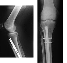
Shin fractures ( fractures ) are common. They are surgically supplied with metal plates and screws or intramedullary nailing through what is known as osteosynthesis .
A common disease caused by overload (jogging, indoor sports, etc.) is anterior tibial edge syndrome ( shin splints ). Occurring in the distal half of the tibia, there is a painful irritation of the fibers of origin of the tibialis anterior muscle .
The Osgood-Schlatter disease is a stimulation of the tibial bump, so the attachment of the patellar ligament. In domestic dogs, a similar disease with detachment of the tibial bump occurs, which is known as tibial tuberosity avulsion .
Rare diseases in children include Jaffé-Campanacci syndrome , tibia recurvata , congenital tibial osteoarthritis , and tibial hemimelia .
literature
- C. Zalpour: Anatomy / Physiology for Physiotherapy. 1st edition. Urban & Fischer, Munich / Jena 2002.
- Franz-Viktor Salomon: Anatomy for veterinary medicine. Enke, Stuttgart 2004, ISBN 3-8304-1007-7 , pp. 37-110.


