Supination trauma
| Classification according to ICD-10 | |
|---|---|
| S93 | Dislocation, sprain, and strain of the joints and ligaments at the level of the upper ankle and foot |
| S93.0 | Dislocation of the upper ankle |
| S93.2 | Traumatic rupture of ligaments at the level of the upper ankle and foot |
| S93.4 | Sprain and strain of the upper ankle |
| ICD-10 online (WHO version 2019) | |
In traumatology , orthopedics and sports medicine, the term supination trauma is understood as the forceful overstretching of the external holding apparatus (joint capsule, ligaments, tendons and bones) of the ankle . The term generally describes the accident-related internal rotation ( supination ) that goes beyond the natural range of motion of a joint , such as is also possible in the wrist or elbow joint (proximal or distal radioulnar joint ), but is used almost exclusively as a synonym for ankle injuries and summarizes this in their different intensities.
Occurrence
The supination trauma is the most common sports injury overall. Grass sports with rapid changes of direction such as ball sports , tennis, badminton and the like are primarily affected, as well as outdoor sports on uneven surfaces such as mountain hiking and climbing (especially when going downhill), jogging, but also Nordic walking. Due to the high to extreme stress on the ankle, these injuries play a special role in dance medicine . Supination trauma is also found in large numbers as an everyday injury (kinking on curbs or steps, etc.).
Accident mechanism
If the foot is stationary, an obstacle or a strong sideways movement can cause the ankle to twist. The ankle bone is turned inwards (“ supinated ”) in relation to the lower leg axis . If the individual resilience of the lateral (outside) holding apparatus is exceeded, its structures can be overstretched or tear.
consequences
Overextension
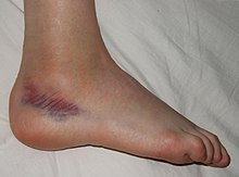
In the simplest form, the capsules and the outer ligaments are only overstretched (" fibular " ligaments: lig. Fibulotalare anterius, lig. Fibulotalare posterius, lig. Fibulocalcaneare), whereby individual fibers of these ligaments and the joint capsule tear, but these structures in their continuity. In this case, we speak of a distortion in the ankle joint. The stability of the joint is not significantly impaired here, but the simultaneous rupture of small blood vessels usually leads to severe pain, swelling and the typical bruise ( hematoma ) below the outer ankle.
Ligament rupture
If one or more of the outer ligaments tear , it is referred to as a fibulotalar ligament rupture ("ligament tear"). In this case, the lateral stability of the upper ankle joint is no longer guaranteed, which is achieved through a load exposure (“held X-ray”) in the form of a “lateral opening” (gaping of the outer joint space) and - if the anterior ligament (lig. Fibulotalare anterius) is involved is - can be documented by the "talus advancement" (displaceability of the ankle bone forwards in relation to the tibial joint surface).
Cartilage or bone damage
Instead of tearing the outer ligament, the tip of the outer ankle can also tear off, in this case the simplest form of ankle joint fracture - the outer ankle fracture type A according to Weber .
Complicating the ligament rupture and the lateral ankle fracture can be a shear injury to the articular cartilage on the ankle bone, a so-called "flake fracture", which can lead to permanent joint damage directly (due to the articular surface defect on the ankle) or indirectly through the cartilage fragment moving in the joint.
The sprained fractures of the upper ankle joint are usually also supination trauma due to their complexity, but are usually not understood under this term and are viewed as an independent injury group.
Diagnosis
A supination trauma can already be suspected from the anamnestic , since the course of the accident is usually described in a typical manner. Clinically, there is painful swelling of the ankle joint, especially in the outer area, often with a hematoma sagging into the connective tissue (bruise, see picture). The ankle joint can be abnormally folded inwards, especially in the case of extensive ligament injuries, if this examination can be expected of the patient due to the pain. X-rays in two directions serve on the one hand to rule out a bony injury, but on the other hand they can also show the ligament instability or the flake fracture that has broken out of the talus. In exceptional cases, further injuries must be excluded or documented with the aid of magnetic resonance imaging .
treatment
Simple distortions are usually adequately treated with elastic bandages and other decongestant measures (cooling, elevation). Ligament ruptures are usually treated functionally - i.e. without immobilizing the joints - with orthotics that reduce the stress on the outer ligaments until they grow together. In exceptional cases (complete rupture of the ligament apparatus with the jamming of ligament or capsule parts in the joint space, flake fracture, external ankle fracture), however, surgical measures may also be necessary. These range from simple ligament sutures to plastic reconstructions of the capsular ligament apparatus to osteosynthetic procedures for external ankle fractures . The duration of treatment depends on the severity of the injury and the individual requirements of the patient (early or delayed start of treatment, compliance , mobility, age, etc.) and can range from a few days to several months.
Long-term consequences
While simple distortions and ligament ruptures usually heal without consequences if treated correctly, accompanying injuries - especially if they remain undetected - occasionally lead to serious long-term consequences:
Ankle arthrosis
Premature wear of the joint surface in the sense of post-traumatic osteoarthritis can occur with injuries to the joint cartilage ("flake fracture") of varying severity; in the long term this can lead to the need for an endoprosthesis (artificial joint ) or arthrodesis (stiffening) of the ankle joint.
Chronic instability
Inadequately treated ligament ruptures can lead to permanent lateral instability of the ankle. Chronic anterolateral rotational instability of the ankle joint due to an inadequately treated and incorrectly healed tear in the anterior fibulotalar ligament and fibulocalcaneal ligament is particularly common . Chronic instability can on the one hand limit the exercise of the preferred sport, on the other hand it can also limit the ability to work. Chronic instability can be depicted with high accuracy using sonography .
To improve balance, exercises for balance and muscle strengthening are used in chronic instability.
Plastic reconstructions of the capsular ligament apparatus may be necessary in the case of chronic instability, but they do not always produce the desired result. In the long term, chronic instability due to incorrect loading of the joint surfaces also leads to (secondary) osteoarthritis, even if there was no primary damage to the articular cartilage.
Individual evidence
- ^ G. Möllenhoff, J. Richter, G. Muhr: The supination trauma - a classic. In: The orthopedist. Volume 28, Number 6, June 1999, pp. 469-475. doi: 10.1007 / PL00003631
- ↑ M. Handschin: Sports injuries on the foot. In: Switzerland Med Forum. 2006; 6, pp. 877-882, PDF
- ↑ Hans Zwipp: Chronic ligament instability of the upper ankle . In: OP-JOURNAL . tape 30 , no. 2 , 2014, p. 104–111 , doi : 10.1055 / s-0034-1383259 ( thieme-connect.de [accessed on November 15, 2018]).
- ^ S. Cao, C. Wang, X. Ma, X. Wang, J. Huang, C. Zhang: Imaging diagnosis for chronic lateral ankle ligament injury: a systemic review with meta-analysis . In: Journal of Orthopedic Surgery and Research . tape 13 , no. 1 , May 2018, p. 122 , doi : 10.1186 / s13018-018-0811-4 , PMID 29788978 , PMC 5964890 (free full text).
- ↑ A. Radwan, J. Bakowski, S. Dew, B. Greenwald, E. Hyde, N. Webber: Effectiveness of ultrasonography in diagnosing lateral ankle instability; A systematic review . In: International Journal of Sports Physical Therapy . tape 11 , no. 2 , April 2016, p. 164-74 , PMID 27104050 , PMC 4827360 (free full text).
- ↑ K. Tsikopoulos, D. Mavridis, D. Georgiannos, MS Cain: Efficacy of non-surgical interventions on dynamic balance in patients with ankle instability: A network meta-analysis . In: Journal of Science and Medicine in Sport . tape 21 , no. 9 , 2018, p. 873-879 , doi : 10.1016 / j.jsams.2018.01.017 , PMID 29571697 .
