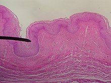
Vaginal epithelium
The vaginal epithelium is the epithelium with which the vagina and the vaginal vestibule are lined, also known as the tunica mucosa vaginae . It is a multilayered, uncornified squamous epithelium. It is rich in glycogen and lies on a lamina propria, which is rich in elastic fibers and wide-lumen veins.
The epithelium is subject during the sexual cycle continuous conversion processes by as sex hormone acting estrogens and progesterone are controlled. These can also be used for diagnosis through cytological examination of a vaginal smear . The vaginal epithelium arises in the embryo from the sinovaginal cusp . In cross-section, the vagina shows the following wall structure:
- Mucous membrane, tunica mucosa vaginae : Determines the surface quality and ensures an acidic environment (pH around 4) through the dead cells of the vaginal epithelium, which, due to the very high glycogen content, represent a good substrate for lactic acid bacteria ( Döderlein bacteria ) and for the development of the special Vaginal flora lead to the microbiome , which counteracts the colonization of bacteria. The actual vaginal epithelium and the underlying lamina propria are included in the tunica mucosa vaginae . The epithelium of the vagina is a multi-layered, uncornified squamous epithelium that can be further differentiated into four layers:
- Basal cell layer , stratum basale
- Parabasal layer , stratum parabasale
- Intermediate layer , stratum intermedium
- Superficial layer , stratum superficiale
They sit on the lamina propria ; it consists of loose connective tissue that is rich in elastic fibers and lymphocytes . In the lamina propria there are capillaries and lymph vessels , from which a transudate is pressed through the epithelium into the vagina during sexual arousal, as well as the plexus venosus vaginalis . The vaginal nerve inversion or sensitivity is minimal. There are only a few free nerve endings; sensory fibers are completely absent.
- Muscle layer, tunica muscularis vaginae : Inside you can see a circular musculature, outside a longitudinal musculature. In this way, the vagina can contract in a ring and lengthways when it enlarges. More precisely, it is smooth muscles embedded in a connective tissue structure . The mesh-like or lattice-like arrangement of the muscle groups has a circular course, also longitudinal on the front wall. The musculature continues in the musculature of the cervix and the perineum. The connective tissue is arranged like a concertina and consists of collagen and numerous elastic fibers. Thanks to this composition, stretching of the vagina is possible.
- Connective tissue layer, tunica adventita vaginae: It contains many elastic fibers and is connected to the connective tissue sheaths of the pelvic floor, urethra and urinary bladder. It is also known as "paracolpium", the dense covering structure made of connective tissue connects the vagina with its surroundings, especially with the urethra. It contains numerous elastic fibers and borders the parametrium cranially.

Individual evidence
- ↑ vaginal epithelium. In: Pschyrembel Medical Dictionary. 257th edition. Walter de Gruyter, Berlin 1993, ISBN 3-933203-04-X , p. 1607.
- ^ Karl Knörr, Henriette Knörr-Gärtner, Fritz K. Beller, Christian Lauritzen: Obstetrics and Gynecology: Physiology and Pathology of Reproduction . 3. Edition. Springer-Verlag, 2013, ISBN 978-3-642-95583-9 , pp. 24-25 .

